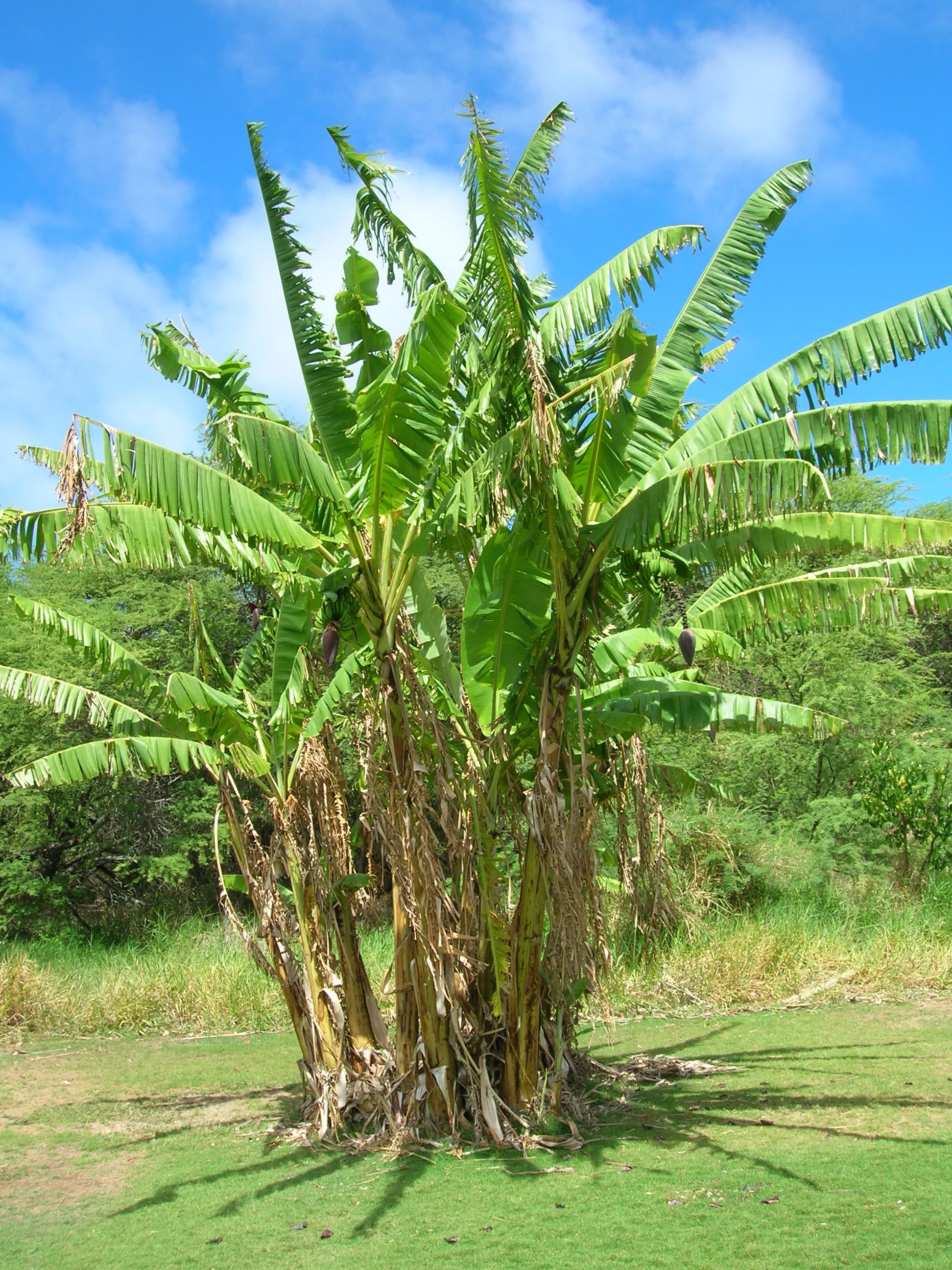
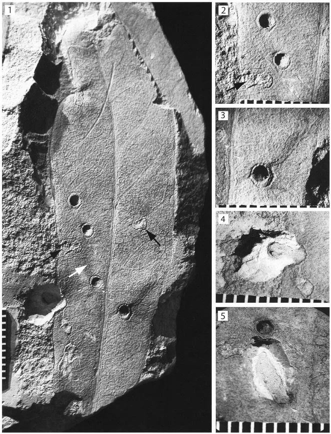
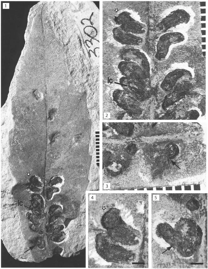
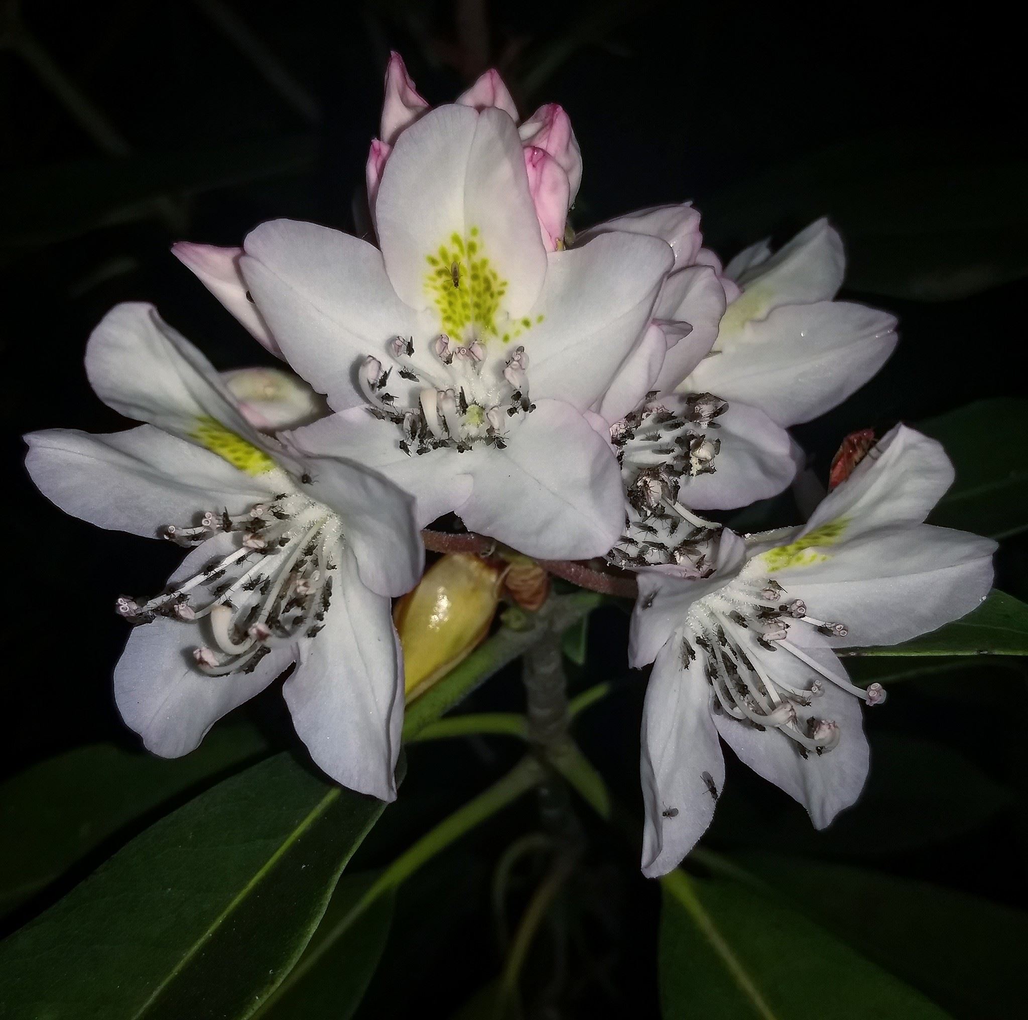
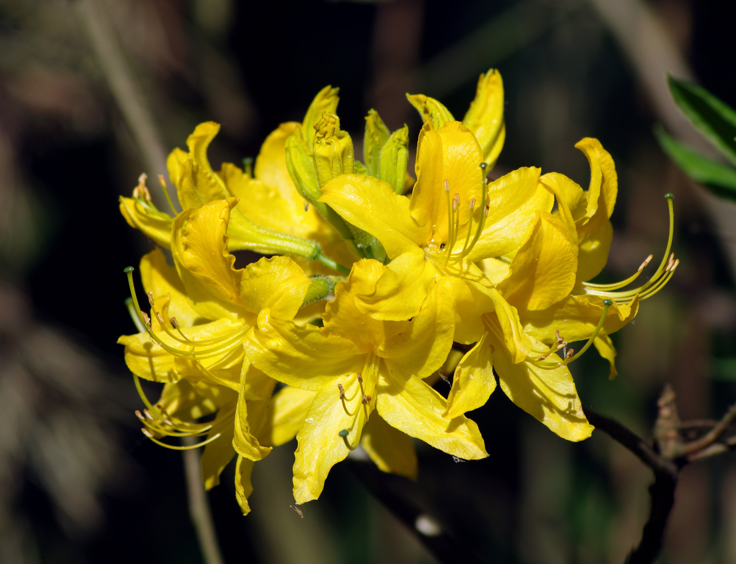
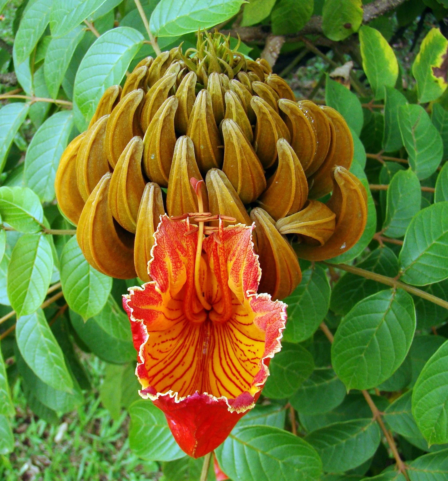
The Role of Leaf Shape on Insect Herbivory

beetle.JPG

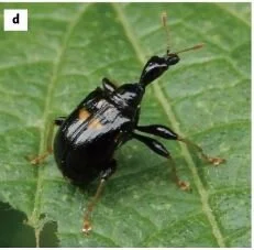
Plants can defend themselves from herbivores in a variety of ways - thorns, spines, hairs, toxins, etc. - but have you ever considered the role of leaf shape in preventing herbivory? It’s okay if you haven’t because leaf shape rarely, if ever, makes it into conversations of plants defense. A recent experiment from Japan has changed that by demonstrating that leaf shape can actually deter a specialist leaf-rolling weevil.

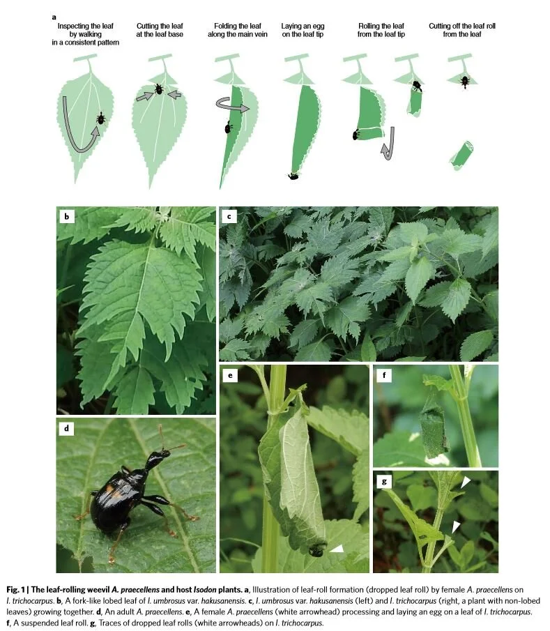
Meet Apoderus praecellens, a leaf rolling weevil that specializes on a genus of mints called Isodon. To successfully reproduce, female leaf rolling weevils must roll up an Isodon leaf while laying eggs as she goes. The end result is a tiny cigar-shaped, edible nursery chamber in which her larvae will develop. The act of processing a leaf is a complex process.

Isodon trichocarpus Photo by Qwert1234 licensed by CC BY-SA 3.0

Isodon trichocarpus Photo by Qwert1234 licensed by CC BY-SA 3.0

The female weevil begins by walking along the margin of the leaf until she reaches the apex. At that point she walks sideways towards the interior of the leaf until she finds the midrib. She then turns around and walks back toward the leaf base again. She repeats these steps several times on both sides of the leaf until she is satisfied. At that point, she will take several bites out of the midrib, which causes the leaf to wilt. The wilted leaf is then much easier to manipulate and thus the rolling process begins.

In the wild, female weevils are well documented on the leaves of I. trichocarpus but not on the leaves of I. umbrosus. This is strange because not only are these plants closely related, they frequently grow in close proximity to one another. Why would the female weevils prefer one over the other? The answer appears to lie in the shape of their leaves.

Isodon trichocarpus produces non-lobed leaves whereas the leaves of I. umbrosus are deeply lobed. When presented with a choice, female weevils did indeed choose to roll I. trichocarpus leaves over those of I. umbrosus. These plants do not differ in their chemical makeup and larvae raised on both species did not differ in their health or development time. Thus, nutritional value or defense compounds don’t explain weevil preference.

leaf roll.JPG

Even more amazing is that the preferences seemed to change when I. trichocarpus leaves were cut to resemble the lobed I. umbrosus leaves. It seems that the presence of leaf lobes is the key to whether a weevil decides to lay her eggs or not. The reason for this seems to be the complex leaf inspection behavior outlined above. The deep lobes of I. umbrosus leaves disrupt the female weevils as they carry out their complex inspection process. If the females are interrupted, they rarely progress to the leaf rolling stage.

The researchers are quick to point out that leaf shape in this instance probably didn’t evolve in response to herbivory. Leaf shape is the result of a multitude of selection pressures like light availability, heat, and drought. Still, the fact that leaf shape can also influence herbivore pressure is an interesting piece to add to the puzzle. It is a great reminder that an organism’s niche comprises so much more than simply the abiotic conditions in which it lives. The niche is also the myriad biological interactions each organism undertakes.

Photo Credits: [1] [2]

Further Reading: [1]

Mutant Orchids Have a lot to Teach Us About Parasitic Plants

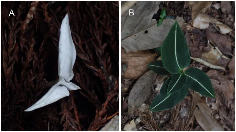
A) Albino and (B) green individual of Goodyera velutina.

A) Albino and (B) green individual of Goodyera velutina.

The botanical world is synonymous with the idea of photosynthesis. Plants take in carbon dioxide and water and utilize light to make their own food. However, not all plants make a living this way. There are many different species of plants that have evolved a parasitic lifestyle to one degree or another. Some of my favorites are those that parasitize mycorrhizal fungi. We call these plants “mycoheterotrophs” and they are fascinating to say the least. Orchids are especially prone to this strategy, with over 1% of all known species having completely lost the ability to photosynthesize.

Our knowledge of the mycoheterotrophic strategy is fragmentary at best. We still don’t fully understand things like how the plants obtain what they need from the fungus nor how they are able to maintain their parasitic lifestyle without the fungus catching on and rejecting the one-sided partnership. This is not to say we know nothing. In fact, as technologies advance, we are unlocking at least some of the mysteries of mycoheterotrophic plants. Some of the best advances come from studying mutant, albino orchids. To understand how, we have to take a closer look at the “average” orchid lifestyle.

Orchids in general make great candidates for understanding the evolution of mycoheterotrophy because all of them start their lives as parasites. Orchids produce some of the smallest seeds in the plant kingdom and without the help of mycorrhizal fungi, they would never be able to germinate. For much of their early life, orchids rely on fungi to provide them with both their mineral and carbohydrate needs. Only after the orchids are large enough to grow leaves will most of them start to give back to their fungal partners in the form of carbohydrates generated from photosynthesis.

Still, many orchids never fully let go of this parasitic lifestyle. This is especially true for orchids living under dense forest canopies. With light in limited supply, many orchids adopt a mixotrophic lifestyle. Essentially this means that although they actively photosynthesize, they nonetheless rely on fungi to provide them with both carbohydrates and minerals. Mixotrphy is likely the most wide-spread orchid strategy and it has been hypothesized that it is also the first step along the path to becoming fully parasitic. This is where the mutant orchids enter the equation.

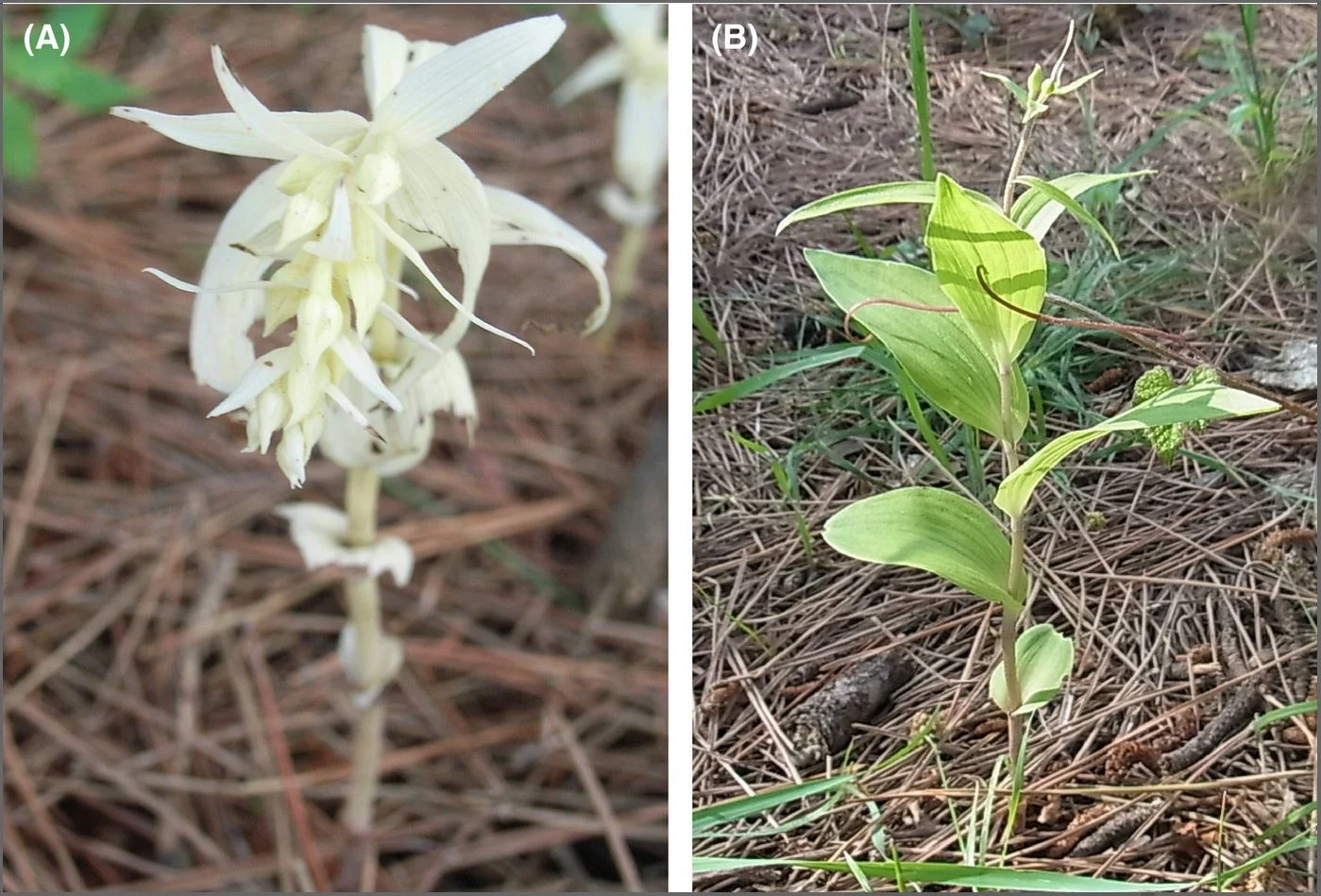
(A) Albino and (B) green individuals of Epipactis helleborine

(A) Albino and (B) green individuals of Epipactis helleborine

Every once in a while, some orchids will germinate and grow into albino versions of their species. Without the ability to produce chlorophyll, these mutants should be destined for a quick death. Such is not the case for many of these orchids. Albino orchids often go on to live full lives, growing and flowering just like their photosynthetic progenitors. Although they do exhibit signs of reduced fitness, the fact that they are able to live at all brings up a lot of questions ready for science to tackle.

Recent investigations into the lives of these albino mutants has revealed some interesting insights into how mycoheterotrophy may have evolved in the first place. By studying the fungal partners of both healthy plants and the albinos, researchers have been able to demonstrate that albinos are doing things a bit differently than their photosynthetic parents. Using isotopes of carbon and nitrogen, scientists are discovering that the albinos have switched their interaction with the fungi in such a way that they more resemble fully mycoheterotrophic orchid species. This is done despite the fact that both albinos and their fully functional parents associate with the same guild of mycorrhizal fungi.

Another interesting difference between albinos and their photosynthetic parents is the fact that the genes involved both antioxidant metabolism and metabolite transfer (mainly carbon in this case) were more active in the albinos than they were in functioning plants. The uptick in gene functioning related to antioxidant metabolism suggests that the mutant plants are undergoing greater oxidative stress than their functional parents. This may have something to do with how the albinos are able to obtain nutrients from their fungal partners. It is thought that mycoheterotrophs actively digest parts of the fungi, which allows them to access the carbon and minerals they need to survive. This process exposes their cells to reactive oxygen compounds that can be very damaging. Antioxidants would help to reduce such damage.

The uptick in genes associated with metabolite transfer was more surprising because it suggests that despite being parasites, the plants are actively transferring substances back to the fungi. It has long been assumed that mycoheterotrophy was a one way street, with fungi transferring nutrients to plants only. These genes now suggest that, at least in some species, such transfer is a two-way street. The exact nature of this two-way transfer remains a mystery and certainly brings up many more questions that must be asked before we can better understand this relationship.

All of this is not to say that such albino mutants are fruitful “next steps” in the evolution of these species. Far from it, in fact. Two things that most albino orchid variants have in common is the fact that they are rare and, of those that have been studied, produce far fewer seeds. There are a lot of anatomical and physiological differences between true mycoheterotrophic species and albino variants and it appears that without those anatomical adaptations, the albinos are a lot less fit than their photosynthetic parents. As authors Selosse and Roy put it:

“non-chlorophyllous variants are likely to represent unique snapshots of failed transitions from mixotrophy to mycoheterotrophy. They are ecological equivalents to mutants in genetics, that is, their dysfunctions might suggest what makes mycoheterotrophy successful. Although their determinism remains unknown, they offer fascinating models for comparing the physiology of mixo- and mycoheterotrophs within similar genetic backgrounds.”

Mutants are strange indeed but with the right kinds of questions and approaches, they have a lot to teach us about ecology, evolution, and life at large.

Photo Credits: [1] [2]

Further Reading: [1] [2] [3]

An Intriguing Way of Presenting One's Pollen

Photo by Monteregina (Nicole) licensed by CC BY-NC-SA 2.0

Photo by Monteregina (Nicole) licensed by CC BY-NC-SA 2.0

Getting pollen from one flower to another is the main reason why flowers exist in the first place. It makes sense then why pollen is often made readily available to pollinators. For many flowering plants, this means directing the pollen-filled anthers outward where they are ready to take advantage of floral visitors. The sunflower family (Asteraceae) does this a bit differently than most. They utilize a technique called secondary pollen presentation.

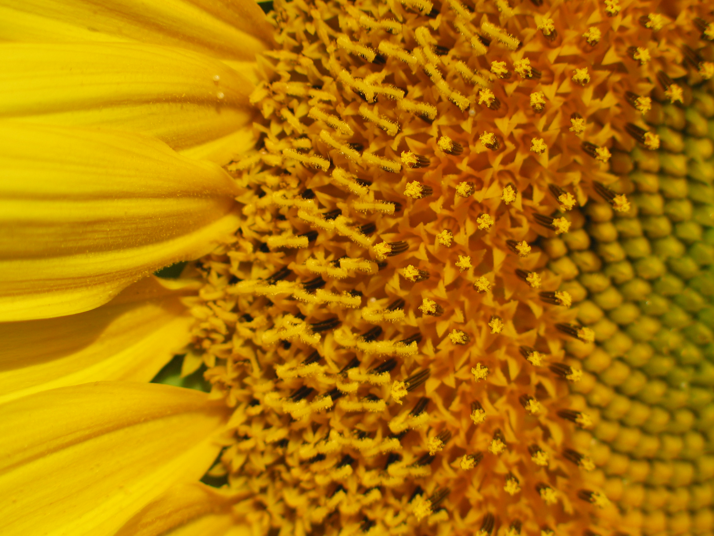
Though secondary pollen presentation is not unique to the sunflower family, their abundance on the landscape makes it super easy to observe. For the sunflower family, what looks like a single flower is actually an inflorescence made up of dense clusters of individual flowers. Each individual flower is roughly tubular in shape and, oddly enough, the anthers are tucked inside the tube facing the interior of the flower. It may seem odd to hide the anthers and their pollen inside of a tube until you see the blooming process sped up.

Photo by László Németh licensed by CC BY-SA 3.0

Photo by László Németh licensed by CC BY-SA 3.0

The sunflower family actually relies on the female parts of the flower to bring the pollen out from the floral tube and into the environment where pollinators can access it. Members of the sunflower family are protandrous, meaning the male parts mature before the female parts. What this means is that the style of the flower can be involved in presenting pollen before it becomes receptive to pollen. This allows enough time for pollen presentation and reduces the likelihood of self pollination.

As the style elongates within the floral tube, one of two things can happen with the pollen inside. In some cases, the style acts like a tiny piston, literally pushing the pollen out into the world. In other cases, the style is covered in tiny, brush-like hairs that rake the pollen from the sides of the floral tube and carry it out as it emerges. In both cases, the style remains closed until enough time has passed for pollen to be taken away from the inflorescence.

Watch _asteraceae GIF on Gfycat. Discover more Timelapse, aster, awesome, back, background, bloom, cool, flower, ground, grow, lapse, out, relax, slender, slow, time, visuals, white, wood, zone GIFs on Gfycat

After a period of time (which varies from species to species), the style splits at the tip and each side curls back on itself to reveal the stigmatic surface. Only at this point in time is are the female parts of the flower mature and ready to receive pollen. With any luck, much of the flowers own pollen would have been collected and taken away to other plants.

The combination of sequential blooming of individual flowers and protandry mean that members of the sunflower family both maximize their chances of pollination and reduce the likelihood of inbreeding. Add to that their ability to disperse their seeds great distances and myriad defense strategies and it should come as no surprise that this family is so darn successful. Get outside and try to witness secondary pollen presentation for yourself. Armed with a hand lens, you will unlock a world of evolutionary wonders!

Photo Credits: [1] [2] [3]

Further Reading: [1] [2]

Surprising Genetic Diversity in Old Growth Trees

Photo by S. Rae licensed by CC BY 2.0

Photo by S. Rae licensed by CC BY 2.0

Long-lived trees face a lot of challenges throughout their lives. Many trees can live for centuries, which can be a problem because plants cannot get up and move when conditions become unfavorable. This should equate to a slower rates of adaptation and evolution for long lived trees but that isn’t always the case. Many trees are often superbly capable of adapting to local conditions. Recently, a team of researchers from the University of British Columbia have provided some insights into the genetic mechanisms that may underpin such adaptive potential.

Genetic insights came from a species of conifer many will be familiar with - the Sitka spruce (Picea sitchensis). Researchers were interested in these trees because they live for a long time (upwards of 500 years or more) and can grow to heights of over 70 meters (230 ft.). They wanted to understand how genetic mutations work in trees like the Sitka spruce because plants are doing things a bit different than animals in that department.

Plants are modular organisms, meaning they grow by producing multiple copies of discrete units. This equates to a branching structure whose overall shape is in large part determined by environmental influences. It also means that when genetic mutations occur in one branch, they can be carried on throughout the growth of those tissues independent of what is going on throughout the rest of the plant. This means that older trees can often accumulate a surprising amount of genetic diversity throughout the entire body of the plant.

Photo by Brandon Kuschel licensed by Creative Commons Attribution 3.0 Unported

Photo by Brandon Kuschel licensed by Creative Commons Attribution 3.0 Unported

When researchers sampled the DNA of tissues from the trunks and the needles of tall, old growth Sitka spruce, they were shocked by what they had found. From the base of the tree to the needles in the canopy, an old growth Sitka spruce can show as much as 100,000 genetic differences. That is a lot of genetic diversity for a single organism. Though plenty of other trees have been found to exhibit varying levels of genetic differences within individuals, this is one of the highest mutation rates ever found in a single eukaryotic organism. This could also explain why such long-lived organisms can survive in a changing world for their entire lives.

Now, it is important to note that many mutations are likely either neutral or potentially harmful. Also, the rates of mutation may differ depending on where you look on this tree. For instance, needles at the top of a Sitka spruce are going to be exposed to far more gene-altering UV radiation than bark tissues near the base. Still, over the lifetime of a single tree, rare beneficial mutations can and do accumulate. Imagine a scenario in which one branch mutation results in needles that are more resistant to say an insect pest. Those needles could hypothetically receive less damage than needles elsewhere on the tree. This odd form of selection is occurring within the lifetime of that tree and may even have implications for the future offspring of that tree thanks again to the quirks of how tree reproductive cells develop.

Many trees also do not have segregated germlines. What this means is that unlike animals whose reproductive cells develop from separate cell lineages than the rest of their body cells, the reproductive cells of trees develop from somatic cells, which are the same cells that form stems, leaves, and branches. This means that if a mutation occurs on the germline of a branch that eventually goes on to produce cones, these mutations can be passed on in the seeds of those cones. This obviously needs a lot of evidence to substantiate but now that a mechanism is in place, we know where and what to look for.

Photo Credits: [1] [2]

Further Reading: [1] [2]

Twinspurs & Their Pollinators

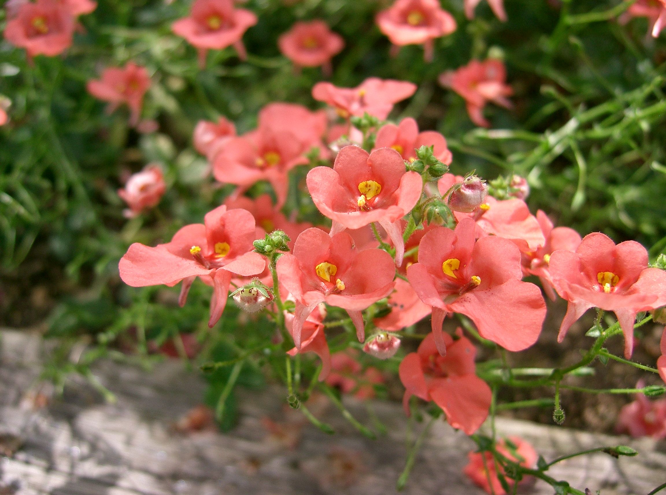
Garden centers and nurseries always have something to teach me. Though I am largely a native plant gardener, the diversity of plant life offered up for sale is always a bit mind boggling. Perusing the shelves and tables of myriad cultivars and varieties, I inevitably encounter something new and interesting to investigate. That is exactly how I came to learn about the twinspurs (Diascia spp.) and their peculiar floral morphology. Far from being simply beautiful, these herbaceous plants have evolved an interesting relationship with a small group of bees.

Diascia whiteheadii. Photo by Ragnhild&Neil Crawford licensed under CC BY-SA 2.0

Diascia whiteheadii. Photo by Ragnhild&Neil Crawford licensed under CC BY-SA 2.0

The genus Diascia comprises roughly 70 species and resides in the family Scrophulariaceae. They are native to a decent chunk of southern Africa and have adapted to a range of climate conditions. Most are annuals but some have evolved a perennial habit. The reason these plants caught my eye was not the bright pinks and oranges of their petals but rather the two spurs that hang off the back of each bloom. Those spurs felt like a bit of a departure from other single-spurred flowers that I am used to so I decided to do some research. I fully expected them to be a mutation that someone had selectively bred into these plants, however, that is not the case. It turns out, those two nectar spurs are completely natural and their function in the pollination ecology of these plants is absolutely fascinating.

Diascia rigescens photo by Dinkum licensed under CC BY-SA 3.0

Diascia rigescens photo by Dinkum licensed under CC BY-SA 3.0

Not all Diascia produce dual spurs on each flower but a majority of them do. The spurs themselves can vary in length from species to species, which has everything to do with their specific pollinator. The inside of each spur is not filled with nectar as one might expect. Instead, the walls are lined with strange trichomes and that secrete an oily substance. It’s this oily substance that is the sole reward for visiting Diascia flowers.

Diascia megathura (a) inflorescenc with arrows indicating spurs and (b) cross sectioned spur showing the trichomes secreting oil (Photos: G. Gerlach).

Diascia megathura (a) inflorescenc with arrows indicating spurs and (b) cross sectioned spur showing the trichomes secreting oil (Photos: G. Gerlach).

If you find yourself looking at insects in southern Africa, you may run into a genus of bees called Rediviva whose females have oddly proportioned legs. The two front legs of Rediviva females are disproportionately long compared to the rest of their legs. They look a bit strange compared to other bees but see one in action and you will quickly understand what is going on. Rediviva bees are the sole pollinators of Diascia flowers. Attracted by the bright colors, the bees alight on the flower and begin probing those two nectar spurs with each of their long front legs.

If you look closely at each front leg, you will notice that they are covered in specialized hairs. Those hairs mop up the oily secretions from within each spur and the bee then transfers the oils to sacs on their hind legs. What is even more amazing is that each flower seems to have entered into a relationship with either a small handful or even a single species of Rediviva bee. That is why the spur lengths differ from species to species - each one caters to the front leg length of each species of Rediviva bee. It is worth noting that at least a few species of Diascia are generalists and are visited by at least a couple different bees. Still, the specificity of this relationship appears to have led to reproductive isolation among many populations of these plants, no doubt lending to the diversity of Diascia species we see today.

Diascia 'Coral Belle' Photo by KENPEI licensed under CC BY-SA 3.0

Diascia 'Coral Belle' Photo by KENPEI licensed under CC BY-SA 3.0

The female bees do not eat the oils they collect. Instead, they take them back to their brood chambers, feed them to their developing offspring, and use what remains to line their nests. At this point it goes without saying that if Diascia were to disappear, so too would these bees. It is incredible to think of the myriad ways that plants have tricked their pollinators into giving up most, if not all of their attention to a single type of flower. Also, I love the fact that a simple trip to a garden center unlocked a whole new world of appreciation for a group of pretty, little bedding plants. It just goes to show you that plants have so much more to offer than just their beauty.

Photo Credits: [1] [2] [3] [4] [5] [6]

Further Reading: [1] [2] [3] [4]

The Wacky World of Whisk Ferns

Photo by Richard Droker licensed under CC BY-NC-ND 2.0

Photo by Richard Droker licensed under CC BY-NC-ND 2.0

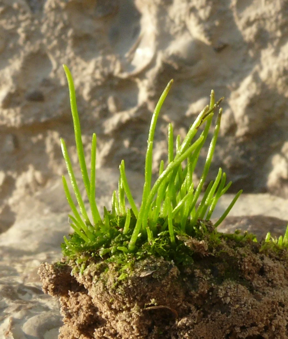
The whisk ferns (Psilotum spp.) are a peculiar group of plants. If you hang out in greenhouses long enough, you are most likely to encounter them as “weeds” growing in pots with other plants. Though they aren’t often put on display by themselves, the whisk ferns are certainly worth a closer look.

Psilotum comprises two species, the far more common Psilotum nudum and the lesser known P. complanatum. These two species will also hybridize, resulting in Psilotum × intermedium. Together, the whisk ferns make up one of only two genera in the family Psilotaceae (the other being Tmesipteris). They are strange plants to look at as there doesn’t appear to be much to them besides stems. Indeed, their peculiar morphology has earned them a fair share of taxonomic attention over the last century but before we get into that, it is a good idea to take a closer look at their anatomy.

Psilotum nudum with yellow sporagia. Photo by Mary Keim licensed under CC BY-NC-SA 2.0

Psilotum nudum with yellow sporagia. Photo by Mary Keim licensed under CC BY-NC-SA 2.0

What we see when we are looking at a whisk fern is the sporophyte generation. Like all sporophytes, its job is to produce the spores that will go on to make new whisk ferns. This part of the whisk fern lifecycle is pretty much all stem. Though these are in fact vascular plants, they do not produce true leaves. Instead, the branching stem takes up all of the photosynthetic work. What looks like tiny leaf-like scales are actually referred to as ‘enations.’ These structures do not contain any vascular tissue of their own. Instead, they bear a type of fused sporangia that house the spores. When mature, these will turn a bright yellow.

Underground, things aren’t much different. Whisk ferns produce a branching rhizome that is covered in hair-like projections called rhizoids. These structures not only help anchor the plant in place, they also function in a similar way to roots. Rhizoids interface with the soil environment allowing the plant to absorb nutrients and water. However, they don’t do this alone. Like so many other plants, whisk ferns partner with mycorrhizal fungi, which vastly increases the amount of surface area these plants have for absorbing what they need. In return, whisk ferns provide the fungi with carbohydrates they produce through photosynthesis. As lovely as this mutualistic relationship sounds, it actually starts off as parasitism.

A Psilotum rhizome with hair-like rhizoids. Photo by Curtis Clark licensed under CC BY-SA 3.0

A Psilotum rhizome with hair-like rhizoids. Photo by Curtis Clark licensed under CC BY-SA 3.0

When the spores find a suitable place to germinate, they will grow into the other half of the whisk fern lifecycle, the gametophyte. These resemble tiny versions of the rhizome and contain male and female reproductive organs. Living underground, the gametophytes do not photosynthesize. Instead, they completely rely on mycorrhizal fungi for all of their nutritional needs. This can go on for some time until the gametophytes are fertilized and grow a new sporophyte. Then and only then will the plant actually start giving back to the fungi that their lives depend on.

Psilotum complanatum with its flattened stems. Photo by Chad Husby licensed under CC BY-NC-ND 2.0

Psilotum complanatum with its flattened stems. Photo by Chad Husby licensed under CC BY-NC-ND 2.0

Because the overall form of the whisk ferns appears so “simplistic.,” many have hypothesized that the genus Psilotum is an evolutionary throwback to the early days of vascular plant evolution. On a superficial level, the whisk ferns do appear to have a lot in common with rhyniophytes, a group of plants that arose during the early Devonian, some 419 to 393 million years ago. A more detailed inspection of the anatomy of each group would reveal that there are some significant and fundamental differences between the two lineages, which I won’t go into here. Also, subsequent molecular work has shown that the whisk ferns reside quite comfortably within the fern lineage and likely represent a sister group to the order that gives us the adder’s tongue ferns (Ophioglossales). It would appear that whisk ferns more accurately represent a reduction in the more “traditional” fern form rather than a holdover from the early days of land plant evolution.

What the genus Psilotum lacks in number of species, it makes up for with its wide distribution. The whisk ferns seem to have conquered most of the tropical and subtropical landmasses on our planet. In fact, I found it incredibly difficult to discern much in the way of a native distribution for these plants. In some areas they are fairly common components of the local flora whereas in others they are considered rare or even threatened. I am sure that at least some of their expansive distribution can be attributed to human assistance as we move soils and plants around the world. To find them in nature, one must look in the cracks of rocks or on the trunks and branches of trees. Though both species can be found growing on trees, P. complanatum in particular seems to prefer an epiphytic lifestyle.

Psilotum complanatum (left) and Psilotum nudum (right) growing epiphytically. Photo by David Eickhoff licensed under CC BY 2.0

Psilotum complanatum (left) and Psilotum nudum (right) growing epiphytically. Photo by David Eickhoff licensed under CC BY 2.0

Whether you grow them on purpose, fight them as a greenhouse “weed,” or track them down in the wild, I hope you take a moment to appreciate these oddball plants. The whisk ferns are intriguing to say the least and certainly offer up a unique conversation piece for anyone curious about the botanical world. They are a genus worth admiring.

Photo Credits: [1] [2] [3] [4] [5]

Further Reading: [1] [2] [3] [4]

A New Case of Lizard Pollination from South Africa

lp1.JPG

With its compact growth habit and small, inconspicuous flowers tucked under its leaves, it seems like Guthriea capensis doesn’t want to be noticed. Indeed, it has earned itself the common name of '“hidden flower.” That’s not to say this plant is unsuccessful. In fact, it seems to do just fine tucked in among high-elevation rock crevices of its home range along the Drakensberg escarpment of South Africa. Despite its cryptic nature, something must be pollinating these plants and recent research has finally figured that out. It appears that the hidden flower has a friend in some local reptiles.

Lizard pollination is not unheard of ([1] & [2]), however, it is by no means a common pollination syndrome. This could have something to do with the fact that we haven’t been looking. Pollination studies are notoriously tricky. Just because something visits a flower does not mean its an effective pollinator. To investigate this properly, one needs ample hours of close observation and some manipulative experiments to get to the bottom of it. Before we get to that, however, its worth getting to know this strange plant in a little more detail.

The hidden flower is a member of an obscure family called Achariaceae. Though a few members have managed to catch our attention economically, most genera are poorly studied. The hidden flower itself appears to be adapted to high elevation environments, hence its compact growth form. By hugging the substrate, this little herb is able to avoid the punishing winds that characterize montane habitats. Plants are dioecious meaning individuals produce either male or female flowers, never both. The most interesting aspect of its flowers, however, are how inconspicuous they are.

The hidden flower (Guthriea capensis) in situ.

The hidden flower (Guthriea capensis) in situ.

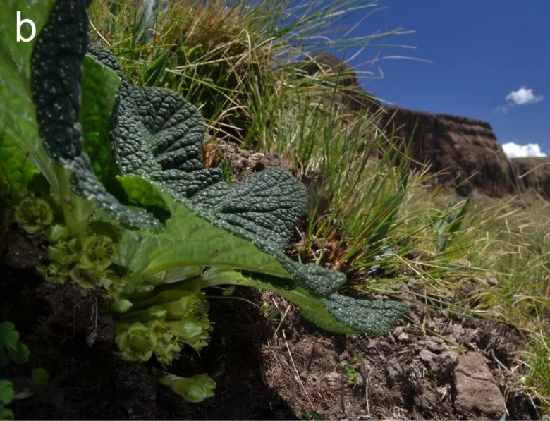
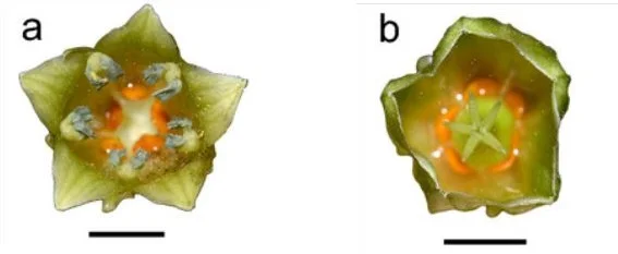
Flowers are produced at the base of the plant, out of site from most organisms. They are small and mostly green in color except for the presence of a few bright orange glands near the base of the style, deep within the floral tube. What they lack in visibility, they make up for in nectar and smell. Each flower produced copious amounts of sticky, sugar-rich nectar. They are also scented. Taken together, these traits usually signal a pollination syndrome with tiny rodents but this assumption appears to be wrong.

Based on hours of video footage and a handful of clever experiments, a team of researchers from the University of KwaZulu-Natal and the University of the Free State have been able to demonstrate that lizards, not mammals, birds, or insects are the main pollinators of this cryptic plant. Two species of lizard native to this region, Pseudocordylus melanotus and Tropidosaura gularis, were the main floral visitors over the duration of the study period.

Pseudocordylus melanotus

Pseudocordylus melanotus

Tropidosaura gularis photo © 2009 Serban Proches licensed under CC BY-SA 2.5

Tropidosaura gularis photo © 2009 Serban Proches licensed under CC BY-SA 2.5

Visiting lizards would spend time lapping up nectar from several flowers before moving off and in doing so, picked up lots of pollen in the process. Being covered in scales means that pollen can have a difficult time sticking to the face of a reptile but the researchers believe that this is where the sticky pollen comes into play. It is clear that the pollen adheres to the lizards’ face thanks to the fact that they are usually covered in sticky nectar. By examining repeated feeding attempts on different flowers, they also observed that not only do the lizards pick up plenty of pollen, they deposit it in just the right spot on the stigma for pollination to be successful. Insect visitors, on the other hand, were not as effective at proper pollen transfer.

Conspicuously absent from the visitation roster were rodents. The reason for this could lie in some of the compounds produced within the nectar. The team found high levels of a chemical called safranal, which is responsible for the smell of the flowers. Safranal is also bitter to the taste and it could very well serve as a deterrent to rodents and shrews. More work will be needed to confirm this hypothesis. Whatever the case, safranal does not seem to deter lizards and may even be the initial cue that lures them to the plant in the first place. Tongue flicking was observed in visiting lizards, which is often associated with finding food in other reptiles.

Male flower (a) and female flower (b). Note the presence of the orange glands at the base.

Male flower (a) and female flower (b). Note the presence of the orange glands at the base.

Another interesting observation is that the color of the floral tube and the orange glands within appear to match the colors of one of the lizard pollinators (Pseudocordylus subviridis ). Is it possible that this is further entices the lizards to visit the flowers? Other reptile pollination systems have demonstrated that lizards appear to respond well to color patterns for which they already have some sort of sensory bias. Is it possible that these flowers evolved in response to such a bias? Again, more work will be needed to say for sure.

By excluding vertebrates from visiting the flowers, the team was able to show that indeed lizards appear to be the main pollinators of these plants. Without pollen transfer, seed set is reduced by 95% wheres the additional exclusion of insects only reduced reproductive success by a further 4%. Taken together, it is clear that lizards are the main pollinators of the enigmatic hidden flower. This discovery expands on our limited knowledge of lizard pollination syndromes and rises many interesting questions about how such relationships evolve.

Photo Credit: [1] [2] [3]

Further Reading: [1] [2]

An Iris With Multiple Parents

DSCN0903.JPG

The Abbeville iris (Iris nelsonii) is a very special plant. It is the rarest of the so-called “Louisiana Irises” and can only be found growing naturally in one small swamp in southern Louisiana. If you are lucky, you can catch it in flower during a few short weeks in spring. The blooms come in a range of colors from reddish-purple to nearly brown, an impressive sight to see siting atop tall, slender stems. However, the most incredible aspect of the biology of this species is its origin. The Abbeville iris is the result of hybridization between not two but three different iris species.

When I found out I would be heading to Louisiana in the spring of 2019, I made sure that seeing the Abbeville iris in person was near the top of my to-do list. How could a botany nut not want to see something so special? Iris nelsonii was only officially described as a species in 1966. Prior to that, many believed hybridization played a role in its origin. Multiple aspects of its anatomy appear intermediate between other native irises. It was not until proper molecular tests were done that the picture became clear.

The Abbeville iris genome contains bits and pieces of three other irises native to Louisiana. The most obvious parent was yet another red-flowering species - the copper iris (Iris fulva). It also contains DNA from the Dixie iris (Iris hexagona) and the zig-zag iris (Iris brevicaulis). If you had a similar childhood as I did, then you may have learned in grade school biology class that hybrids are usually biological dead ends. They may exhibit lots of beneficial traits but, like mules, they are often sterile. Certainly this is often the case, especially for hybrid animals, however, more and more we are finding that hybridization has resulted in multiple legitimate speciation events, especially in plants.

Iris fulva. Photo by Richard licensed under CC BY-NC-ND 2.0

Iris fulva. Photo by Richard licensed under CC BY-NC-ND 2.0

Iris hexagona. Photo by beautifulcataya licensed under CC BY-NC-ND 2.0

Iris hexagona. Photo by beautifulcataya licensed under CC BY-NC-ND 2.0

Iris brevicaulis. Photo by peganum licensed under CC BY-SA 2.0

Iris brevicaulis. Photo by peganum licensed under CC BY-SA 2.0

How exactly three species of iris managed to “come together” and produce a functional species like I. nelsonii is interesting to ponder. Each of its three parent species prefers a different sort of habitat than the others. For instance, the copper iris is most often found in seasonally wet, shady bottomland hardwood forests as well as the occasional roadside ditch, whereas the Dixie iris is said to prefer more open habitats like wet prairies. In a few very specific locations, however, these types of habitats can be found within relatively short distances of each other.

Apparently at some point in the past, a few populations swapped pollen and the eventual result was a stable hybrid that would some day be named Iris nelsonii. As mentioned, this is a rare plant. Until it was introduced to other sites to ensure its ongoing existence in the wild, the Abbeville iris was only known to occur in any significant numbers at one single locality. This necessitates the question as to whether or not this “species” is truly unique in its ecology to warrant that status. It could very well be that that single locality just happens to produce a lot of one off hybrids.

In reality, the Abbeville iris does seem to “behave” differently from any of its parental stock. For starters, it seems to perform best in habitats that are intermediate of its parental species. This alone has managed to isolate it enough to keep the Abbeville from being reabsorbed genetically by subsequent back-crossing with its parents. Another mechanism of isolation has to do with its pollinators. The Abbeville iris is intermediate in its floral morphology as well, which means that pollen placement may not readily occur when pollinators visit different iris species in succession. Also, being largely red in coloration, the Abbeville iris receives a lot of attention from hummingbirds.

Although hummingbirds do not appear to show an initial preference when given the option to visit copper and Abbeville irises at a given location, research has found that once hummingbirds visit an Abbeville iris flower, they tend to stick to that species provided enough flowers are available. As such, the Abbeville iris likely gets the bulk of the attention from local hummingbirds while it is in bloom, ensuring that its pollen is being delivered to members of its own species and not any of its progenitors. For all intents and purposes, it would appear that this hybrid iris is behaving much like a true species.

As with any rare plant, its ongoing survival in the wild is always cause for concern. Certainly Louisiana is no stranger to habitat loss and an ever-increasing human population coupled with climate change are ongoing threats to the Abbeville iris. Changes in the natural hydrologic cycle of its swampy habitat appear to have already caused a shift in its distribution. Whereas it historically could be found in abundance in the interior of the swamp, reductions in water levels have seen it move out of the swamp and into ditches where water levels remain a bit more stable year round. Also, if its habitat were to become more fragmented, the reproductive barriers that have maintained this unique species may degrade to the point in which it is absorbed back into an unstable hybrid mix with one or a couple of its parent species. Luckily for the Abbeville, offspring have been planted into at least one other location, which helps to reduce the likelihood of extinction due to a single isolated event.

Further Reading: [1] [2] [3] [4] [5]

The Succulent Passionflowers

Photo by Wendy Cutler licensed by CC BY-SA 2.0

Photo by Wendy Cutler licensed by CC BY-SA 2.0

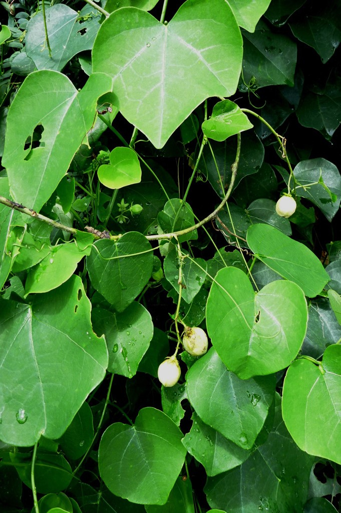
Succulent passionflowers?! It took me a minute to get my head wrapped around the idea. It wasn’t until I saw one in flower that I truly understood. The genus Adenia is found throughout east and west Africa, Southeast Asia, and hits its peak diversity in Madagascar. It comprises approximately 100 species and, as a whole, is poorly understood. Today I would like to introduce you to this bizarre genus within Passifloraceae.

Adenia glauca Photo by Karelj licensed under the GNU Free Documentation License

Adenia glauca Photo by Karelj licensed under the GNU Free Documentation License

Adenia is, to date, the second largest genus within the Passionflower family and yet delineating species has been something of a nightmare for botanists over the years. At least some of this confusion lies within the diversity of this odd group. It has been said that few angiosperm lineages surpass Adenia in the diversity of growth forms they exhibit. Though all could be considered succulent to some degree, Adenia runs the gamut from trees to vines, and even tuberous herbs.

3505817218_274303366e_o.jpg

Even within individual species, the overall form of these plants can vary widely depending on the conditions under which they have been growing. Their succulent nature and that fact that many species can reach rather large proportions means that herbarium records for this group are scant at best. Many are only known from a single, incomplete collection of a few bits and pieces of plant. Also, juvenile plants often look very different from their adult forms, making timing of the collection crucial for proper analysis.

To complicate matters more, all Adenia are dioecious, meaning that individual plants are either male or female. Male and female flowers of individual species look pretty distinct and differ a bit from what we have come to expect out of the passionflower family. Often collections were made on only a single sex. This is further complicated by the fact that these plants often exhibit very short flowering seasons. Most come into bloom right before the onset of the rainy season and are entirely leafless at that point in time. Because of this, it has been extremely difficult to accurately match flowering collections to vegetative collections. As such, nearly 1/4 of all Adenia species are missing descriptions of either male or female flowers and their fruits.

Female flower of Adenia reticulata. Photo by C. E. Timothy Paine licensed under CC BY-NC 2.0

Female flower of Adenia reticulata. Photo by C. E. Timothy Paine licensed under CC BY-NC 2.0

Male flowers of Adenia digitata. Photo by Joachim Beyenbach licensed under CC BY-SA 3.0

Male flowers of Adenia digitata. Photo by Joachim Beyenbach licensed under CC BY-SA 3.0

Flowers of Adenia firingalavensis.  Photo by voyage-madagascar.org licensed under CC BY 2.0

Flowers of Adenia firingalavensis. Photo by voyage-madagascar.org licensed under CC BY 2.0

Fruits of Adenia hondala

Fruits of Adenia hondala

Even genetic work has failed to clear up much of the mysteries that surround this group. Some studies suggest that Adenia is sister to all other genera within Passifloraceae whereas others have even suggested it to be nestled neatly within the genus Passiflora. The most recent work hints at a placement among the tribe Passifloreae. If this confuses you, you are certainly not alone. Until a more complete sampling effort is done on Adenia, I think it is safe to say that this genus will be holding onto its taxonomic mysteries for the foreseeable future.

Adenia globosa photo by KENPEI licensed under the GNU Free Documentation License

Adenia globosa photo by KENPEI licensed under the GNU Free Documentation License

All Adenia are perennial plants but how they manage this differs from species to species. Some put all of their energy into underground tubers, producing annual stems and leaves that die back each year. Others don’t produce any tubers and instead store all of their water and nutrients within thick stems. This has made at least a handful of species a hit with succulent growers around the world. It is always an interesting sight to see a giant caudiciform trunk or base with bunches of spindly stems spraying out from the top.

Leaves and fruit of Adenia cissampeloides. Photo by International Institute of Tropical Agriculture licensed under CC BY-NC 2.0

Leaves and fruit of Adenia cissampeloides. Photo by International Institute of Tropical Agriculture licensed under CC BY-NC 2.0

Juvenile Adenia glauca.  Photo by laurent houmeau licensed under CC BY-SA 2.0

Juvenile Adenia glauca. Photo by laurent houmeau licensed under CC BY-SA 2.0

Adenia are also extremely toxic plants. The conditions under which these plants evolved are tough and it appears that this group doesn’t want to take any chances on losing any biomass to herbivores. The main class of compounds they produce are called lectins. These proteins cause myriad issues within animal bodies including rapid cell death, blood clotting, inhibition of protein synthesis, and a disruption of ribosome and DNA function. Needless to say, its in any critters best interest to avoid nibbling on any species of Adenia. Even handling and pruning of these plants merits caution.

Photo by Wendy Cutler licensed under CC BY 2.0

Photo by Wendy Cutler licensed under CC BY 2.0

Whether you’re a botanist, taxonomist, gardener, or just curious about plant diversity, Adenia is a wonderful example of just how many unknowns are still out there. Regardless of their taxonomic status, these are fascinating species, each with a wonderful ecology and intriguing evolutionary history. These plants are hardy survivors and a great example of the lengths a genus can go to when presented with new opportunities. Undoubtedly many more species await description but the plants we currently know of are fascinating to say the least.

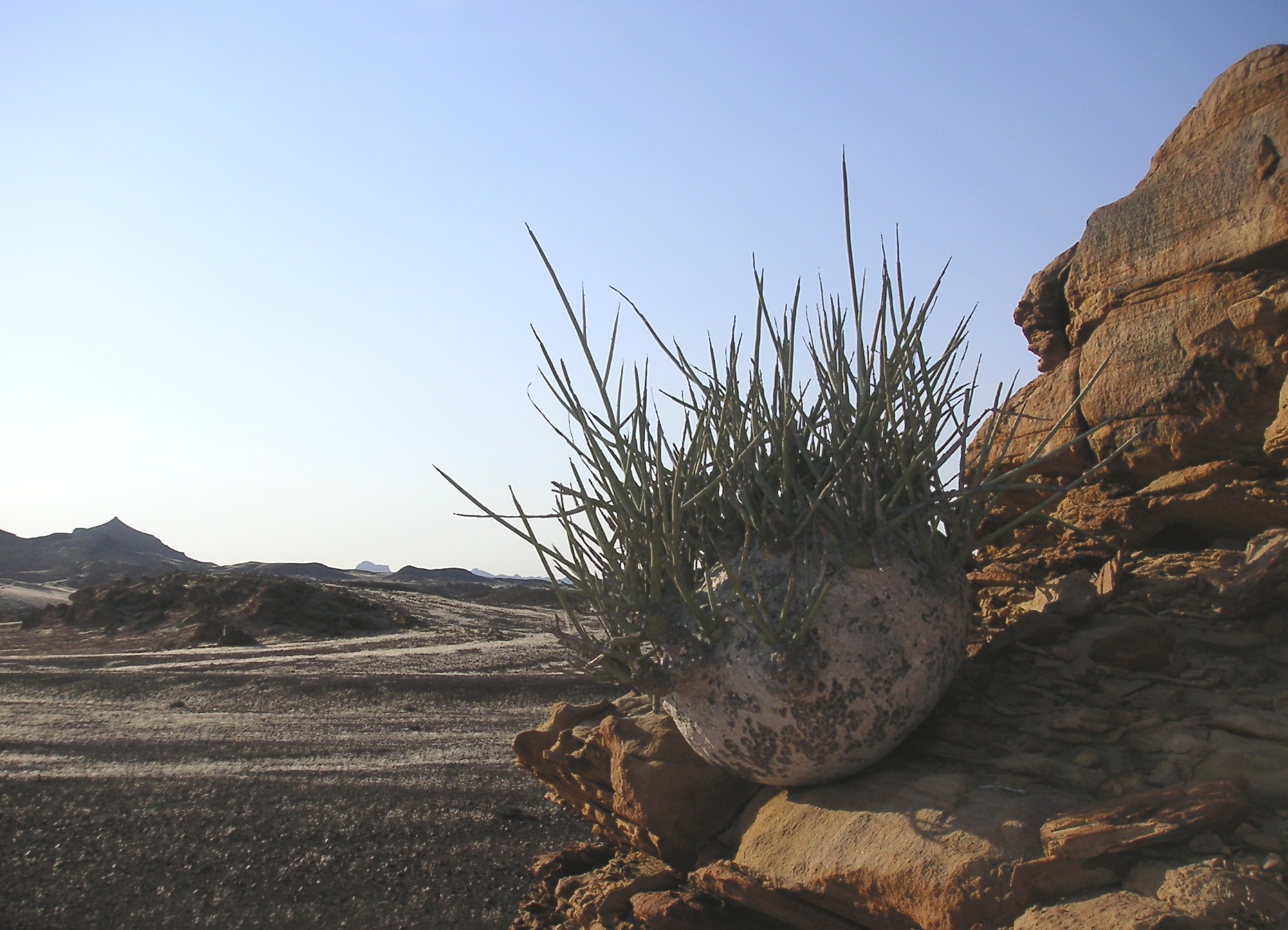
Adenia pechuelii. Photo by Ewald Schmidt licensed under public domain.

Adenia pechuelii. Photo by Ewald Schmidt licensed under public domain.

Photo Credits: [1] [2] [3] [4] [5] [6] [7] [8] [9] [10] [11] [12] [13]

Further Reading: [1] [2]

The Drought Alert System of Terrestrial Plants has an Underwater Origin

Photo by Christian Fischer licensed by CC BY-SA 3.0

Photo by Christian Fischer licensed by CC BY-SA 3.0

For plants, the transition from water to land was a monumental achievement that changed our world forever. Such a transition was fraught with unique challenges, not the least of which being the ever present threat of desiccation. A new study now suggests that those early land plants already had the the tools to deal with drought and they have their aquatic algal ancestors to thank.

One of the keys to being able to survive drought is being able to detect it in the first place. Without some sort of signalling pathway, plants would not be able to close up stomata and channel vital water and nutrients to more important tissues and organs. As such, elucidating the origins and function of drought signalling pathways in plants has been of great interest to science.

One key set of pathways involved in plant drought response is collectively referred to as the “chloroplast retrograde signaling network.” I’m not even going to pretend that I understand how these pathways operate in any detail but there is one aspect of this network that is the key to this recent discovery. It involves the means by which drought and high-light conditions are sensed in one part of the plant and how that information is then communicated to the rest of the plant. When this signalling pathway is activated, the plant can then begin to produce enzymes that go on to activate defense strategies such as stomatal closure.

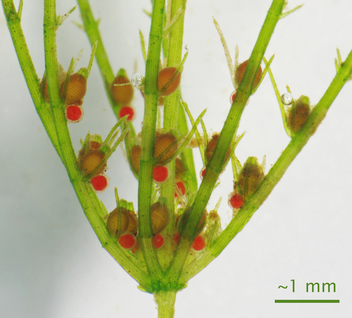
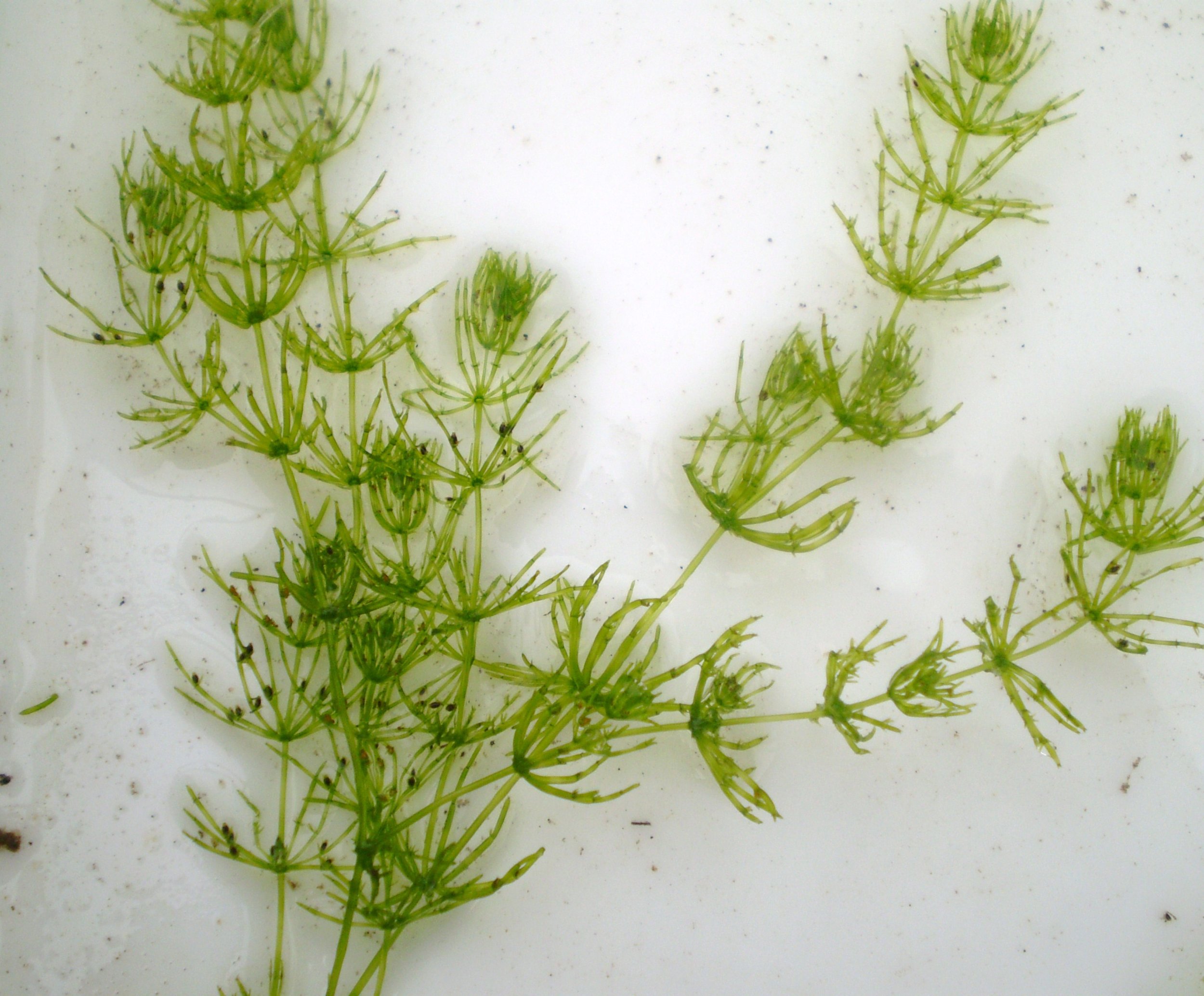
Chara braunii - a modern day example of a streptophyte alga. Photo by Show_ryu licensed under the GNU Free Documentation License

Chara braunii - a modern day example of a streptophyte alga. Photo by Show_ryu licensed under the GNU Free Documentation License

The surprise came when researchers at the Australian National University, in collaboration with researchers at the University of Florida, decided to study the chloroplast retrograde signaling network in more detail. They were interested in the inner workings of this process in relation to stomata. Stomata are tiny pores on the leaves and stems of terrestrial plants that regulate the exchange of gases like CO2 and oxygen as well as water vapor. To add some controls to their experiment, the team added a few species of aquatic algae into the mix. Algae do not produce stomata and therefore they reasoned that no traces of chloroplast retrograde signaling network enzymes should be present.

This is not what happened. Instead, the team discovered that the enzymes in question also showed up in a group of algae known as the streptophytes. This was exciting because streptophyte algae hail from the lineage thought to be ancestral to all land plants. It appears that the tools necessary for terrestrial plants to survive drought were already in place before their ancestors ever left the water.

Why this is the case could have something to do with the streptophyte lifestyle. Today, these algae are known to tolerate very tough conditions. Though outright drought is rarely a threat for these aquatic algae, they nonetheless have to deal with scenarios that resemble drought such as high salinity. Streptophyte algae found growing in ephemeral pools must cope with ever increasing concentrations of salinity as the water around them evaporates. It is possible that this drought signalling pathway may have evolved as a response to hyper-saline conditions such as these. Regardless of what was going on during those early days of plant evolution, this research indicates that the ability for terrestrial plants to deal with drought evolved before their ancestors ever left the water.

The closer we look, the more we can appreciate that evolution of important traits isn’t always de novo. More often it appears that new innovations result from a retooling of of older genetic equipment. In the case of land plants, a signalling pathway that allowed their aquatic ancestors to deal with water loss was coopted later on by organs such as leaves and stems to deal with the stresses of life on land. As the old saying goes, “life uhhh… finds a way.”

Photo Credits: [1] [2]

Further Reading: [1] [2]
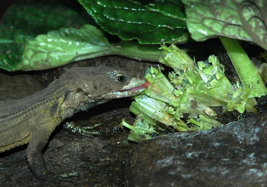
Süßwassertang: A Fern Disguised as a Liverwort

Photo by Rǫgn licensed under CC BY-SA 4.0

Photo by Rǫgn licensed under CC BY-SA 4.0

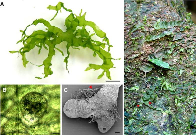
If you enjoy planted aquariums, you may have crossed paths with a peculiar little plant called Süßwassertang. It can be propagated by breaking off tiny pieces, which eventually grow into a tangled carpet of tiny green thalli. One could be excused for thinking that Süßwassertang was some sort of liverwort and indeed, for quite some time was marketed as such. That all changed in 2009 when it was revealed that this was not a liverwort at all but rather the gametophyte of a fern.

Despite its German name, Süßwassertang appears to have originated in tropical parts of Africa and Asia. It is surprisingly hard to find out any information about this plant outside of its use in the aquarium trade. The name Süßwassertang translates to “freshwater seaweed” and indeed, that is exactly what it looks like. The fact that this is actually the gametophyte of a fern may seem startling at first but when you consider what they must deal with in nature, the situation makes a bit more sense.

A Süßwassertang gametophyte. B An antheridium, showing a cap cell (cc), ring cell (rc), and basal cell (bc). Bar: 20 µm. C Developing lateral branches with rhizoids (arrowhead) and meristems (m) Bar: 0.2 mm. D Ribbon-like, branched gametophyte (g) o…

A Süßwassertang gametophyte. B An antheridium, showing a cap cell (cc), ring cell (rc), and basal cell (bc). Bar: 20 µm. C Developing lateral branches with rhizoids (arrowhead) and meristems (m) Bar: 0.2 mm. D Ribbon-like, branched gametophyte (g) of L. spectabilis bearing a young sporophyte (sp) Bar: 1 cm

Fern gametophytes are surprisingly hardy considering their small size and delicate appearance. They are amazing in their ability to tolerate harsh conditions like drought and freezing temperatures. Because of this, fern gametophytes sometimes establish themselves in places that would be unfavorable for their sporophyte generation. For some, this means never completing their lifecycle. Others, however, seem to have overcome the issue by remaining in their gametophyte stage forever. Though no sexual reproduction occurs for these permanent gametophytes, they nonetheless persist and reproduce by breaking off tiny pieces, which grow into new colonies.

The sporophyte of a related species, Lomariopsis marginata, demonstrating the usual epiphytic habit of this genus. Photo by Alex Popovkin, Bahia, Brazil licensed under CC BY-NC-SA 2.0

The sporophyte of a related species, Lomariopsis marginata, demonstrating the usual epiphytic habit of this genus. Photo by Alex Popovkin, Bahia, Brazil licensed under CC BY-NC-SA 2.0

This appears to be the case for Süßwassertang. Amazingly, despite a few attempts, no sporophytes have ever been coaxed from any gametophyte. It would appear that this is yet another species that has given up its sporophyte phase for an entirely vegetative habit. What is most remarkable is what the molecular work says about Süßwassertang taxonomically. It appears that this plant its nestled into a group of epiphytic ferns in the genus Lomariopsis. How this species evolved from vine-like ferns living in trees to an asexual colony of aquatic gametophytes is anyones’ guess but it is an incredible jump to say the least.

Photo Credits: [1] [2] [3]

Further Reading: [1]
Gooey Pitcher Fluids

Photo by Shawn Mayes licensed under CC BY-SA 3.0

Photo by Shawn Mayes licensed under CC BY-SA 3.0

There seems to be no end to the diversity of colors, shapes, and sizes exhibited by Nepenthes and their pitchers. These wonderful carnivorous plants grow these pitchers as a means of supplementing their nutritional needs as the habitats in which Nepenthes are found are lacking in vital nutrients like nitrogen. There are as many variations on the pitcher theme as there are Nepenthes but all function as traps in one form or another. How they trap insects is another topic entirely and some species have evolved incredible means of making sure prey does not escape. Some of my favorites belong to those species that employ sticky mucilage.

Arguably one of the most iconic of this type is Nepenthes inermis. This species is endemic to a small region of Sumatra and, to date, has only been found growing on a handful of mountain peaks in the western part of the country. The specific epithet ‘inermis’ is Latin for ‘unarmed’ as was given in reference to the bizarre upper pitchers of this plant. They look more like toilet bowls than anything carnivorous and indeed, they lack many of the features characteristic of other Nepenthes pitchers such as a peristome and a slippery, waxy coating on the inside of the pitcher walls.

Photo by Alfindra Primaldhi licensed under CC BY 2.0

Photo by Alfindra Primaldhi licensed under CC BY 2.0

These may seem like minor details but consider the role these features play in other Nepenthes. A peristome is essentially a brightly colored, slippery lip that lines the outer rim of the pitcher mouth. Not only does this serve in attracting insect prey, it also aids in their capture. As mentioned, the peristome can be extremely slippery (especially when wet) so that any insect stumbling around on the rim is much more likely to fall in. Once inside, a waxy coating on the inside of some pitchers aids in keeping insects down. They simply cannot get purchase on the waxy walls and therefore cannot climb back out. So, for N. inermis to lack both features is a bit strange.

Another interesting feature of N. inermis pitchers is the highly reduced pitcher lid. It hasn’t disappeared completely but compared with other Nepenthes, this pitcher lid barely registers as one. For most Nepenthes, pitcher lids serve multiple functions. For starters, they keep the rain out. Nepenthes are most at home in humid, tropical climates where rain is a daily force to be reckoned with. For many Nepenthes, rain not only dilutes the valuable digestive soup brewing within each pitcher, it can also cause them to overflow and dump their nutritious contents. Pitcher lids can also help in attracting prey. Like the peristome, they are often brightly colored but many also secrete nectar, which insects find irresistible. Lured in by the promise of food, some insects inevitably fall down into the pitcher below.

Looking into the pitcher of Nepenthes inermis. Photo by Shawn Mayes licensed under CC BY-SA 3.0

Looking into the pitcher of Nepenthes inermis. Photo by Shawn Mayes licensed under CC BY-SA 3.0

Considering the importance of such structures, it becomes a little bit confusing why some Nepenthes have evolved away from this anatomy. The question then remains, why would a species like N. inermis no longer produce pitchers with these features? Amazingly, the answer actually lies within the pitcher fluid itself.

Tip over the upper pitchers of N. inermis and you will soon discover that they are filled with an extremely viscous mucilage. It is so viscous that some have reported that when the pitchers are held upside down, the mucilage within can form an unbroken stream of considerable length. Its the viscosity of this fluid that is the real reason that N. inermis is able to capture prey so easily. Insects lured to the traps can catch a drink of the nectar on the tiny lid. In doing so, some inevitably fall down into the pitcher itself.

The upper pitcher of the closely related Nepenthes dubia. Photo  by Alfindra Primaldhi licensed under CC BY 2.0

The upper pitcher of the closely related Nepenthes dubia. Photo by Alfindra Primaldhi licensed under CC BY 2.0

Instead of slippery walls or downward pointing hairs keeping the insects in, the viscous pitcher fluid quickly engulfs the struggling prey. Some have even suggested that the nectar secreted by the tiny lid has narcotic effects on visiting insects, however, I have not seen any data demonstrating this. Once caught in the fluid, insects easily slide their way down into the depths of the pitcher where they can be digested. This is probably why the pitchers are shaped like tiny toilet bowls; their shape allows for a large sticky surface area for insects to get stuck while prey that has already been captured is funneled down to where digestion and absorption takes place. In a way, these types of pitchers behave surprisngly similar to the sticky traps utilized by other carnivorous plants like sundews (Drosera spp.).

The viscous fluid also comes in handy during the frequent rains that blanket these mountains. As mentioned above, rain would quickly dilute most pitcher fluids but not when the pitcher fluid itself is more dense. Water sits on top of the viscous mucilage and when the pitchers become too heavy, they tip over. The water readily pours out but little if any of the pitcher fluid is lost in the process. It seems that species like N. inermis no longer fight the elements but rather have adapted to meet them head on. As such, they no longer have a need for a large pitcher lid.

Nepenthes jamban takes the toilet bowl shape to the extreme. Photo  by Alfindra Primaldhi licensed under CC BY 3.0

Nepenthes jamban takes the toilet bowl shape to the extreme. Photo by Alfindra Primaldhi licensed under CC BY 3.0

Nepenthes inermis is not alone in having evolved pitchers like this. Viscous pitcher mucilage is a trait shared by its closest relatives - N. dubia, N. flava, N. jacquelineae, N. jamban, N. talangensis, and N. tenuis, as well as even more distantly related species such as N. rafflesiana. Because prey capture is so important for the fitness of individuals, it is no wonder that so many different forms have evolved within this genus. In fact, many experts believe that variations in the way in which prey is captured and utilized is one of the main reasons why Nepenthes have undergone such a dramatic adaptive radiation.

Sadly, the uniqueness in form and function of these pitchers has landed many of these species on the endangered species list. As if habitat destruction wasn’t already pushing some to the brink, species like N. inermis are being poached at alarmingly unsustainable rates. Due to their limited distributions, most populations simply cannot recover from even moderate levels of harvesting. The silver lining in all of this is that many Nepenthes are extremely easy to grow and propagate provided their basic needs are met. As more and more folks enter into the carnivorous plant hobby, hopefully more and more people will be sharing seeds, cuttings, and tissue cultured materials. In doing so, we can hopefully reduce some of the pressures placed on wild populations.

Photos via Wikimedia Commons

Further Reading: [1] [2] [3]

An Introduction to Hornworts

Anthoceros sp. Photo by Bramadi Arya licensed under CC BY-SA 4.0

Anthoceros sp. Photo by Bramadi Arya licensed under CC BY-SA 4.0

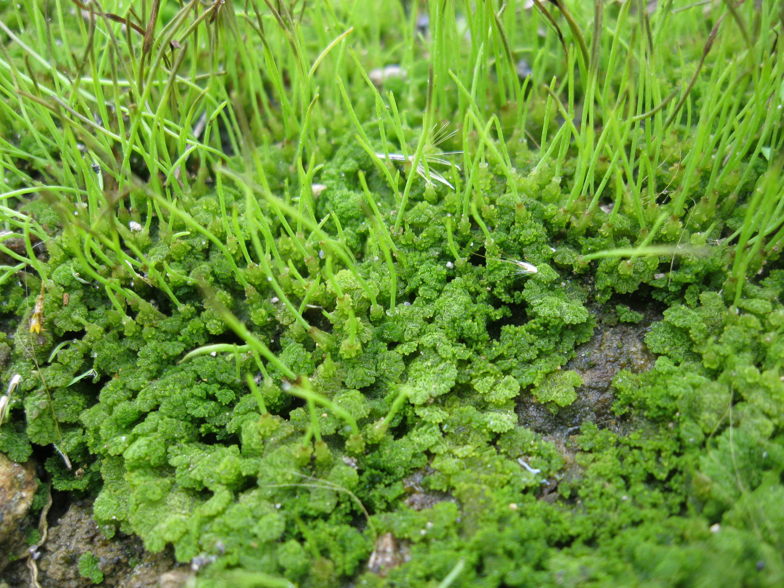
When was the last time you thought about hornworts? Have you ever thought about hornworts? If you answered no, you aren’t alone. Despite their global distribution, these tiny plants receive hardly any attention and that is a shame. Hornworts (Anthocerotophyta) have been around for a very long time. In fact, it is likely that they were some of the first plants to colonize the land roughly 300 - 400 million years ago.

To be fair, hornworts aren’t known for their size. They are generally small plants, though their colonies can form impressive mats. To find them, one must try looking in and among rocks, bare patches of soil, or pretty much anywhere enough moisture builds up to supply their needs. They tend to enjoy nutrient-poor substrates but I would hesitate to say that with any certainty. No matter where you live, from the tundra to the tropics, there is probably a hornwort native to your neck of the woods.

Dendroceros sp. Photo by J.Ziffer licensed under public domain

Dendroceros sp. Photo by J.Ziffer licensed under public domain

How many different species of hornwort there are is apparently the subject of some debate. Some authors recognize upwards of 300 species whereas others suggest the real number hangs somewhere around 150. Regardless of the exact numbers, hornworts belong to one of six genera: Anthoceros, Dendroceros, Folioceros, Megaceros, Notothylas and Phaeoceros. Fun fact, the suffix ‘ceros’ at the end of each genus is derived from the Latin word for ‘horn.’

The reason they are called hornworts is because of their reproductive structures or “sporophytes.” Similar to their moss and liverwort cousins, hornworts undergo an alternation of generations in order to reproduce sexually. The green gametophytes house the sexual organs - antheridia if they are male and archegonia if they are female. After fertilization, a sporophyte begins to grow, which will go on to produce and disseminate spores. However, the way in which the hornwort sporophyte forms is a bit different from what we see in mosses and liverworts.

Alternation of generations in hornworts. Photo by Mariana Ruiz (LadyofHats) licensed under public domain

Alternation of generations in hornworts. Photo by Mariana Ruiz (LadyofHats) licensed under public domain

Upon fertilization, the zygote begins to divide into a bulbous mass of cells affectionately referred to as "the foot.” This foot remains within the gametophyte throughout the lifetime of the hornwort, depending on the gametophyte for water and nutrients. Even more peculiar is the the fact that the growing point of the sporophyte is at the base rather than the tip. As such, the horn of each hornwort could continue to grow upwards until it is damaged in some way.

The horn itself is an amazing structure. Whereas the outside layers of tissue are merely structural, the internal tissues differentiate into two different types - spores and pseudo-elaters. Pseudo-elaters expand and contract as humidity fluctuates so as the sporophyte splits to release the spores, the pseudo-elaters dehydrate and snap like tiny spore catapults, thus aiding in their dispersal.

Megaceros flagellaris. Photo by Dr. Scott Zona licensed under CC BY-NC 2.0

Megaceros flagellaris. Photo by Dr. Scott Zona licensed under CC BY-NC 2.0

Of course, reproduction is the main goal but to get to that point, hornworts must grow and mature. How they manage to survive is incredible because it is a reminder that what are often thought of as “primitive” plants are actually far more advanced than we give them credit for. The main body of the hornwort gametophyte is a thin layer of cells that spread out to form a tiny, green mat. This is the structure you are most likely to encounter.

Inside each cell is a single chloroplast. In most hornworts, the chloroplast does not exist in isolation. Instead, it is fused with other organelles into a structure called a “pyrenoid.” The pyrenoid functions as both a center for photosynthesis and a food storage organ. This is unique as it relates to terrestrial plants but quite common in algae. Another odd fact about hornwort anatomy are the presence of tiny cavities scattered throughout their tissues. These cavities form as clusters of hornwort cells die. They then fill with a special mucilage that appears to invite colonization by nitrogen-fixing cyanobacteria. The cyanobacteria set up shop within the cavities and provides the hornwort with supplemental nitrogen in return for a place to live.

Anthoceros agrestis photo by BerndH licensed under CC BY-SA 3.0

Anthoceros agrestis photo by BerndH licensed under CC BY-SA 3.0

Cyanobacteria aren’t the only organisms to have partnered with hornworts either. Mycorrhizal fungi also enter into the picture. A study done back in 2013 actually found that a wide variety of fungi will partner with hornworts which suggests that this symbiotic relationship is much more ancient and versatile than we once thought. Fungi cluster around parts of the gametophyte that produce root-like structures called “rhizoids,” offering nutrients in return for carbohydrates.

All in all, I think it is safe to say that hornworts are remarkable little plants. Though they can sometimes be difficult to find and properly identify, they nonetheless offer plenty of inspiration for the botanically inclined mind. We can all do better by tiny plants like the hornworts. They have been on land for an incredible amount of time and they definitely deserve our respect and admiration.

Photo Credits: [1] [2] [3] [4] [5]

Further Reading: [1] [2] [3]

The Rise and Fall of the Scale Trees

Photo by Ghedoghedo licensed under CC BY-SA 3.0

Photo by Ghedoghedo licensed under CC BY-SA 3.0

If I had a time machine, the first place I would visit would be the Carboniferous. Spanning from 358.9 to 298.9 million years ago, this was a strange time in Earth’s history. The continents were jumbled together into two great landmasses - Laurasia to the north and Gondwana to the south and the equatorial regions were dominated by humid, tropical swamps. To explore these swamps would be to explore one of the most alien landscapes this world has ever known.

The Carboniferous was the heyday for early land plants. Giant lycopods, ferns, and horsetails formed the backbone of terrestrial ecosystems. By far the most abundant plants during these times were a group of giant, tree-like lycopsids known as the scale trees. Scale trees collectively make up the extinct genus Lepidodendron and despite constantly being compared to modern day club mosses (Lycopodiopsida), experts believe they were more closely related to the quillworts (Isoetopsida).

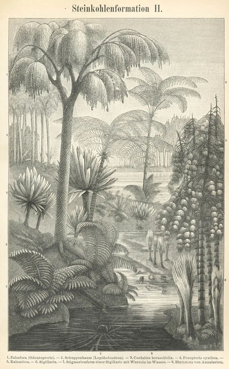
Carboniferous coal swamp reconstruction dating back to the 1800’s

Carboniferous coal swamp reconstruction dating back to the 1800’s

It is hard to say for sure just how many species of scale tree there were. Early on, each fragmentary fossil was given its own unique taxonomic classification; a branch was considered to be one species while a root fragment was considered to be another, and juvenile tree fossils were classified differently than adults. As more complete specimens were unearthed, a better picture of scale tree diversity started to emerge. Today I can find references to anywhere between 4 and 13 named species of scale tree and surely more await discovery. What we can say for sure is that scale tree biology was bizarre.

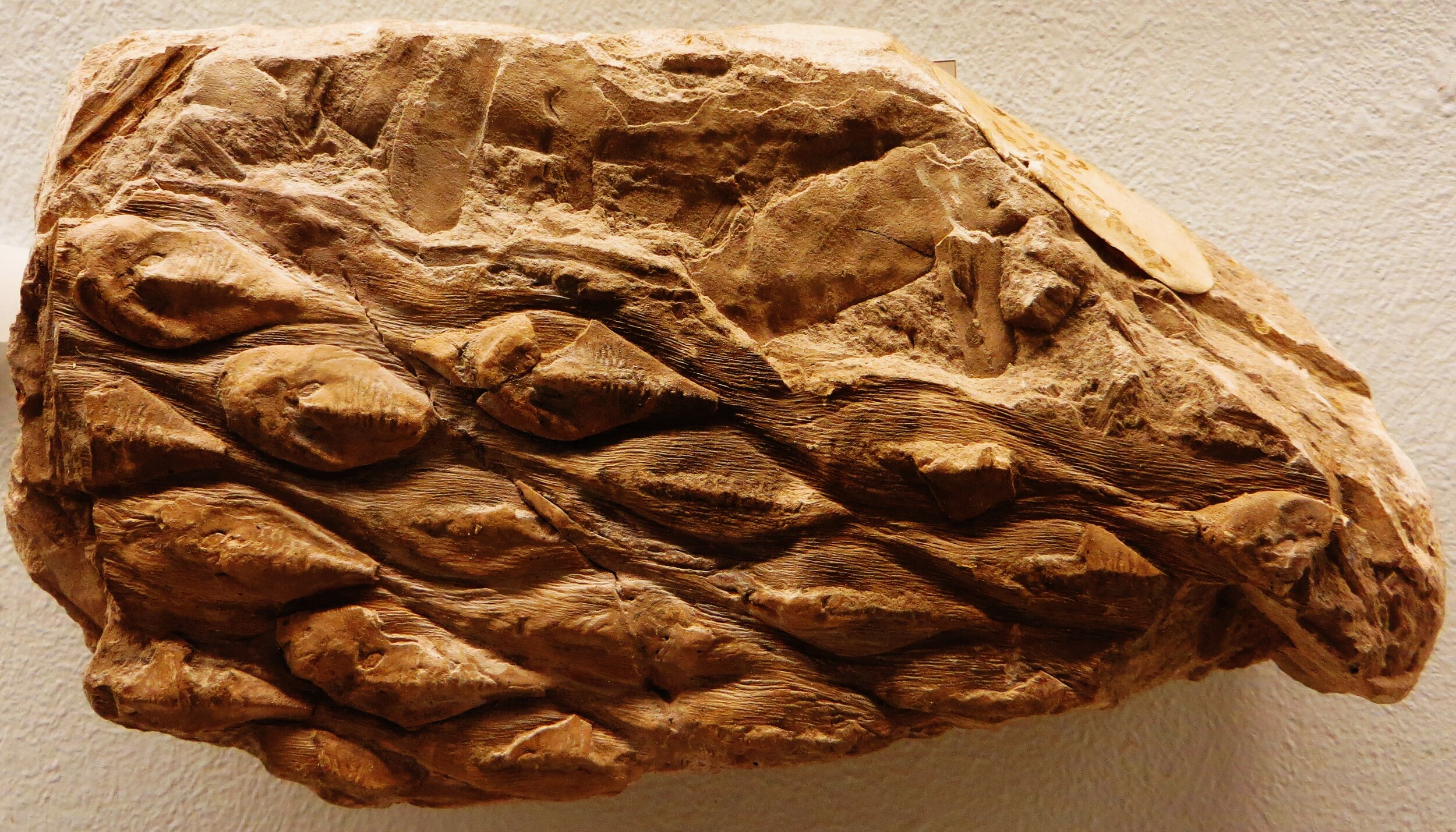
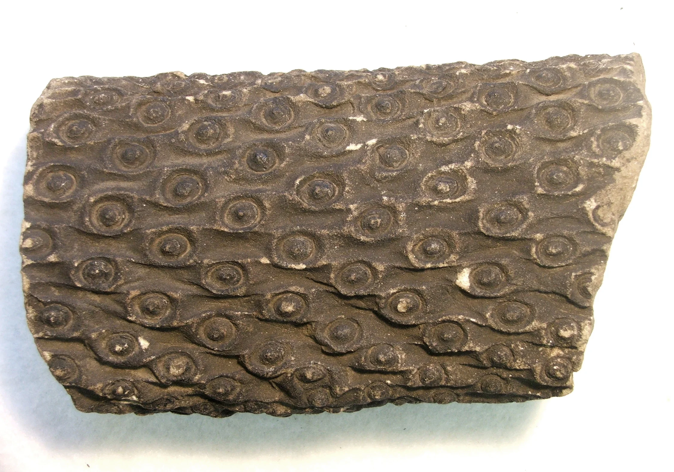
The name “scale tree” stems from the fossilized remains of their bark, which resembles reptile skin more than it does anything botanical. Fossilized trunk and stem casts are adorned with diamond shaped impressions arranged in rows of ascending spirals. These are not scales, of course, but rather they are leaf scars. In life, scale trees were adorned with long, needle-like leaves, each with a single vein for plumbing. Before they started branching, young trees would have resembled a bushy, green bottle brush.

As scale trees grew, it is likely that they shed their lower leaves, which left behind the characteristic diamond patterns that make their fossils so recognizable. How these plants achieved growth is rather fascinating. Scale tree cambium was unifacial, meaning it only produced cells towards its interior, not in both directions as we see in modern trees. As such, only secondary xylem was produced. Overall, scale trees would not have been very woody plants. Most of the interior of the trunk and stems was comprised of a spongy cortical meristem. Because of this, the structural integrity of the plant relied on the thick outer “bark.” Many paleobotanists believe that this anatomical quirk made scale trees vulnerable to high winds.

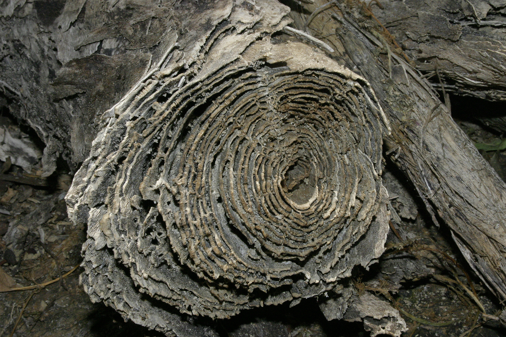
Scale trees were anchored into their peaty substrate by rather peculiar roots. Originally described as a separate species, the roots of these trees still retain their species name. Paleobotanists refer to them as “stigmaria” and they were unlike most roots we encounter today. Stigmaria were large, limb-like structures that branched dichotomously in the soil. Each main branch was covered in tiny spots that were also arranged in rows of ascending spirals. At each spot, a rootlet would have grown outward, likely partnering with mycorrhizal fungi in search of water and nutrients.

A preserved Lepidodendron stump

A preserved Lepidodendron stump

Eventually scale trees would reach a height in which branching began. Their tree-like canopy was also the result of dichotomous branching of each new stem. Amazingly, the scale tree canopy reached staggering heights. Some specimens have been found that were an estimated 100 ft (30 m) tall! It was once thought that scale trees reached these lofty heights in as little as 10 to 15 years, which is absolutely bonkers to think about. However, more recent estimates have cast doubt on these numbers. The authors of one paper suggest that there is no biological mechanism available that could explain such rapid growth rates, concluding that the life span of a typical scale tree was more likely measured in centuries rather than years.

Regardless of how long it took them to reach such heights, they nonetheless would have been impressive sites. Remarkably, enough of these trees have been preserved in situ that we can actually get a sense for how these swampy habitats would have been structured. Whenever preserved stumps have been found, paleobotanists remark on the density of their stems. Scale trees did not seem to suffer much from overcrowding.

leps.PNG

The fact that they spent most of their life as a single, unbranched stem may have allowed for more success in such dense situations. In fact, those that have been lucky enough to explore these fossilized forests often comment on how similar their structure seems compared to modern day cypress swamps. It appears that warm, water-logged conditions present similar selection pressures today as they did 350+ million years ago.

Like all living things, scale trees eventually had to reproduce. From the tips of their dichotomosly branching stems emerged spore-bearing cones. The fact that they emerge from the growing tips of the branches suggests that each scale tree only got one shot at reproduction. Again, analyses of some fossilized scale tree forests suggests that these plants were monocarpic, meaning each plant died after a single reproductive event. In fact, fossilized remains of a scale tree forest in Illinois suggests that mass reproductive events may have been the standard for at least some species. Scale trees would all have established at around the same time, grown up together, and then reproduced and died en masse. Their death would have cleared the way for their developing offspring. What an experience that must have been for any insect flying around these ancient swamps.

The fossilized strobilus of a Lepidodendron. Photo by Verisimilus T licensed under the GNU Free Documentation License.

The fossilized strobilus of a Lepidodendron. Photo by Verisimilus T licensed under the GNU Free Documentation License.

Compared to modern day angiosperms, the habits of the various scale trees may seem a bit inefficient. Nonetheless, this was an extremely successful lineage of plants. Scale trees were the dominant players of the warm, humid, equatorial swamps. However, their dominance on the landscape may have actually been their downfall. In fact, scale trees may have helped bring about an ice age that marked the end of the Carboniferous.

You see, while plants were busy experimenting with building ever taller, more complex anatomies using compounds such as cellulose and lignin, the fungal communities of that time had not yet figured out how to digest them. As these trees grew into 100 ft monsters and died, more and more carbon was being tied up in plant tissues that simply weren’t decomposing. This lack of decomposition is why we humans have had so much Carboniferous coal available to us. It also meant that tons of CO2, a potent greenhouse gas, were being pulled out of the atmosphere millennia after millennia.

A fossilized root or “stigmaria”. Photo by Verisimilus T licensed under CC BY-SA 3.0

A fossilized root or “stigmaria”. Photo by Verisimilus T licensed under CC BY-SA 3.0

As atmospheric CO2 levels plummeted and continents continued to shift, the climate was growing more and more seasonal. This was bad news for the scale trees. All evidence suggests that they were not capable of keeping up with the changes that they themselves had a big part in bringing about. By the end of the Carboniferous, Earth had dipped into an ice age. Earth’s new climate regime appeared to be too much for the scale trees to handle and they were driven to extinction. The world they left behind was primed and ready for new players. The Permian would see a whole new set of plants take over the land and would set the stage for even more terrestrial life to explode onto the scene.

It is amazing to think that we owe much of our industrialized society to scale trees whose leaves captured CO2 and turned it into usable carbon so many millions of years ago. It seems oddly fitting that, thanks to us, scale trees are once again changing Earth’s climate. As we continue to pump Carboniferous CO2 into our atmosphere, one must stop to ask themselves which dominant organisms are most at risk from all of this recent climate change?

Photo Credits: [1] [2] [4] [5] [6] [7]

Further Reading: [1] [2] [3] [4] [5] [6] [7] [8] [9] [10] [11] [12] [13] [14]
Gymnosperms and Fleshy "Fruits"

Fleshy red aril surrounding the seeds of Taxus baccata. Photo by Frank Vincentz licensed under the GNU Free Documentation License.

Fleshy red aril surrounding the seeds of Taxus baccata. Photo by Frank Vincentz licensed under the GNU Free Documentation License.

Many of us were taught in school that one of the key distinguishing features between gymnosperms and angiosperms is the production of fruit. Fruit, by definition, is a structure formed from the ovary of a flowering plant. Gymnosperms, on the other hand, do not enclose their ovules in ovaries. Instead, their unfertilized ovules are exposed (to one degree or another) to the environment. The word “gymnosperm” reflects this as it is Greek for “naked seed.” However, as is the case with all things biological, there are exceptions to nearly every rule. There are gymnosperms on this planet that produce structures that function quite similar to fruits.

Internal anatomy of a Ginkgo ovule with red arrow showing the integument.Photo copyright Bruce Kirchoff, Licensed under CC BY 2.0

Internal anatomy of a Ginkgo ovule with red arrow showing the integument.

Photo copyright Bruce Kirchoff, Licensed under CC BY 2.0

The key to understanding this evolutionary convergence lies in understanding the benefits of fruits in the first place. Fruits are all about packing seeds into structures that appeal to the palates of various types of animals who then eat said fruits. Once consumed, the animals digest the fruity bits and will often deposit the seeds elsewhere in their feces. Propagule dispersal is key to the success of plants as it allows them to not only to complete their reproductive cycle but also conquer new territory in the process. With a basic introduction out of the way, let’s get back to gymnosperms.

“Fruits” of Cephalotaxus fortunei (Cephalotaxaceae)

“Fruits” of Cephalotaxus fortunei (Cephalotaxaceae)

There are 4 major gymnosperm lineages on this planet - the Ginkgo, cycads, gnetophytes, and conifers. Each one of these groups contains members that produce fleshy structures around their seeds. However, their “fruits” do not all develop in the same way. The most remarkable thing to me is that, from a developmental standpoint, each lineage has evolved its own pathway for “fruit” production.

Ginkgo “fruits” are full of butyric acid and smell like rotting butter or vomit. Photo by H. Zell licensed under CC BY-SA 3.0

Ginkgo “fruits” are full of butyric acid and smell like rotting butter or vomit. Photo by H. Zell licensed under CC BY-SA 3.0

For instance, consider ginkgos and cycads. Both of these groups can trace their evolutionary history back to the early Permian, some 270 - 280 million years ago, long before flowering plants came onto the scene. Both surround their developing seed with a layer of protective tissue called the integument. As the seed develops, the integument swells and becomes quite fleshy. In the case of Ginkgo, the integument is rich in a compound called butyric acid, which give them their characteristic rotten butter smell. No one can say for sure who this nasty odor originally evolved to attract but it likely has something to do with seed dispersal. Modern day carnivores seem to be especially fond of Ginkgo “fruits,” which would suggest that some bygone carnivore may have been the main seed disperser for these trees.

“Fruits” contained within the female cone of a cycad (Lepidozamia peroffskyana). Photo by Tony Rodd licensed under CC BY-NC-SA 2.0

“Fruits” contained within the female cone of a cycad (Lepidozamia peroffskyana). Photo by Tony Rodd licensed under CC BY-NC-SA 2.0

The Gnetophytes are represented by three extant lineages (Gnetaceae, Welwitschiaceae, and Ephedraceae), but only two of them - Gnetaceae and Ephedraceae - produce fruit-like structures. As if the overall appearance of the various Gnetum species didn’t make you question your assumptions of what a gymnosperm should look like, its seeds certainly will. They are downright berry-like!

Berry-like seeds of Gnetum gnemon. Photo by gbohne licensed under CC BY-SA 2.0

Berry-like seeds of Gnetum gnemon. Photo by gbohne licensed under CC BY-SA 2.0

The formation of the fruit-like structure surrounding each seed can be traced back to tiny bracts at the base of the ovule. After fertilization, these bracts grow up and around the seed and swell to become red and fleshy. As you can imagine, Gnetum “fruits” are a real hit with animals. In the case of some Ephedra, the “fruit” is also derived from much larger bracts that surround the ovule. These bracts are more leaf-like at the start than those of their Gnetum cousins but their development and function is much the same.

Red, fleshy bracts of Ephedra distachya. Photo by Le.Loup.Gris licensed under CC BY-SA 3.0

Red, fleshy bracts of Ephedra distachya. Photo by Le.Loup.Gris licensed under CC BY-SA 3.0

Whereas we usually think of woody cones when we think of conifers, there are many species within this lineage that also have converged on fleshy structures surrounding their seeds. Probably the most famous and widely recognized example of this can be seen in the yews (Taxus spp.). Ovules are presented singly and each is subtended by a small stalk called a peduncle. Once fertilized, a group of cells on the peduncle begin to grow and differentiate. They gradually swell and engulf the seed, forming a bright red, fleshy structure called an “aril.” Arils are magnificent seed dispersal devices as birds absolutely relish them. The seed within is quite toxic so it usually escapes the process unharmed and with any luck is deposited far away from the parent plant.

The berry-like cones of Juniperus communis. Photo by Piero Amorati, ICCroce - Casalecchio di Reno, Bugwood.org licensed under Creative Commons Attribution 3.0 License.

The berry-like cones of Juniperus communis. Photo by Piero Amorati, ICCroce - Casalecchio di Reno, Bugwood.org licensed under Creative Commons Attribution 3.0 License.

Another great example of fleshy conifer “fruits” can be seen in the junipers (Juniperus spp.). Unlike the other gymnosperms mentioned here, the junipers do produce cones. However, unlike pine cones, the scales of juniper cones do not open to release the seeds inside. Instead, they swell shut and each scale becomes quite fleshy. Juniper cones aren’t red like we have seen in other lineages but they certainly garnish the attention of many a small animal looking for food.

I have only begun to scratch the surface of the fruit-like structures in gymnosperms. There is plenty of literary fodder out there for those of you who love to read about developmental biology and evolution. It is a fascinating world to uncover. More importantly, I think the fleshy “fruits” of the various gymnosperm lineages stand as a testament to the power of natural selection as a driving force for evolution on our planet. It is amazing that such distantly related plants have converged on similar seed dispersal mechanisms by so many different means.

Photo Credits: [1] [2] [3] [4] [5] [6] [7] [8]

Further Reading: [1] [2] [3] [4] [5] [6] [7]
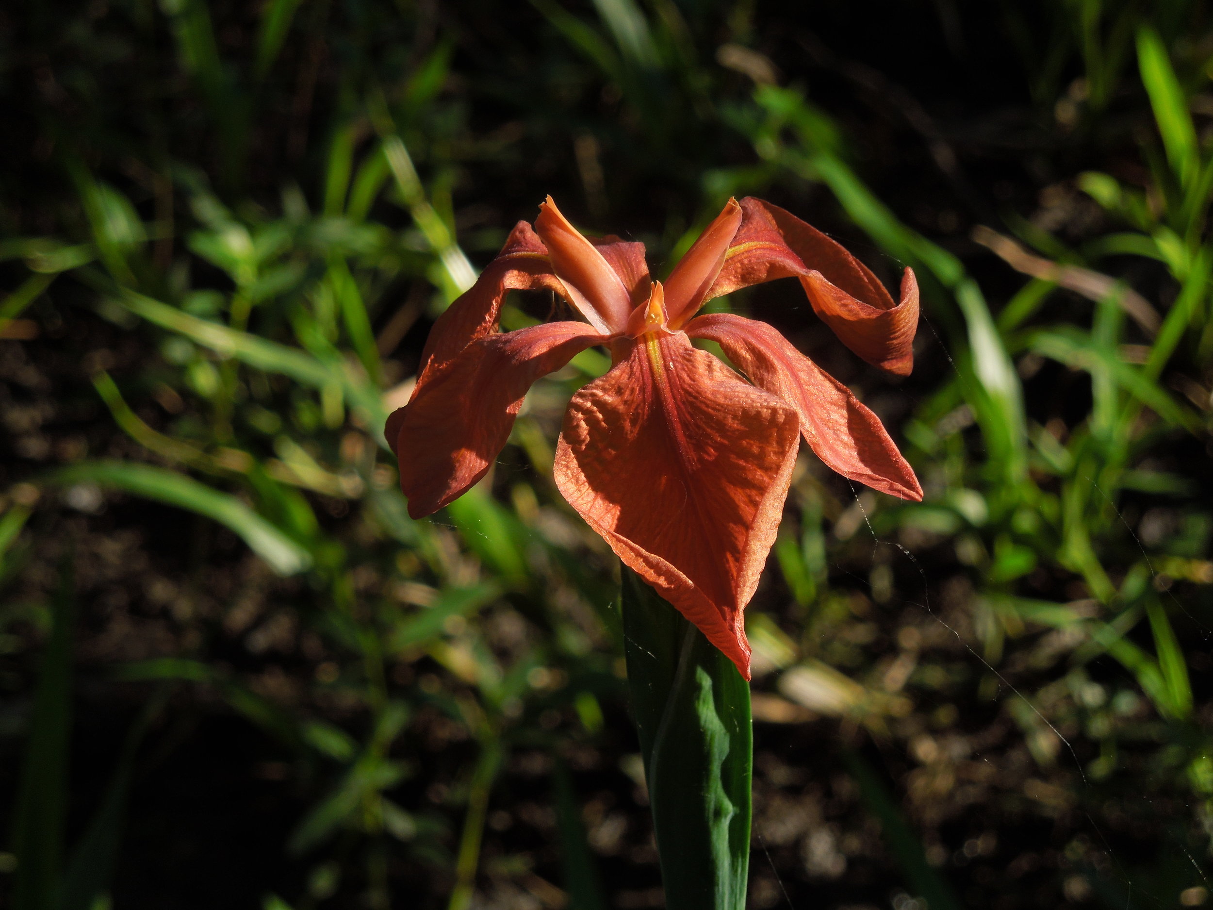
Meet Pokeweed's Tree-Like Cousin

Photo by Roberto Fiadone licensed under CC BY-SA 4.0

Photo by Roberto Fiadone licensed under CC BY-SA 4.0

There is more than one way to build a tree. For that reason and more, “tree” is not a taxonomic designation. Arborescence has evolved independently throughout the botanical world and many herbaceous plants have tree-like relatives. I was shocked to learn recently of a plant native to the Pampa region of South America commonly referred to as ombú. At first glance it looks like some sort of fig, with its smooth bark and sinuous, buttressed roots. Deeper investigation revealed that this was not a fig. Ombú is actually an arborescent cousin of pokeweed!

Photo by Dick Culbert licensed under CC BY 2.0

Photo by Dick Culbert licensed under CC BY 2.0

The scientific name of ombú is Phytolacca dioica. As its specific epithet suggests, plants are dioecious meaning individuals are either male or female. Unlike its smaller, herbaceous cousins, ombú is an evergreen perennial. Because they can grow all year, these plants can reach bewildering proportions. Heights upwards of 60 ft. (18 m.) are not unheard of and the crowns of more robust specimens can easily attain diameters of 40 to 50 ft. (12 - 18 m.)! What makes such sizes all the more impressive is the way in which ombú is able to achieve such growth.

Photo by Lanntaron licensed under CC BY-SA 3.0

Photo by Lanntaron licensed under CC BY-SA 3.0

Ombú is thought to have evolved from an herbaceous ancestor. Cut into the trunk of one of these trees and you will see that this phylogenetic history has left its mark. Ombú do not produce what we think of as wood. Instead, much of the support for branches and stems comes from turgor pressure. Also, the way in which these trees grow is not akin to what you would see from something like an oak or a maple. Whereas woody trees undergo secondary growth in which the cambium layer differentiates into xylem and phloem, thus thickening stems and roots, ombú exhibits a unique form of stem and root thickening called “anomalous secondary thickening.”

Essentially what this means is that instead of a single layer of cambium forming xylem and phloem, ombú cambium exhibits unidirectional thickening of the cambium layer. There are a lot of nitty gritty details to this kind of growth and I must admit I don’t have a firm grasp on the mechanics of it all. The point of the matter is that anomalous secondary thickening does not produce wood as we know it and instead leads to rapid growth of weak and spongy tissues. This is why turgor pressure is so important to the structural integrity of these trees. It has been estimated that the trunk and branches of an ombú is 80% water.

A cross section of an ombú limb showing harder xylem tissues separated by spongy parenchyma that has since disintegrated. Photo by Tony Rodd licensed under CC BY-NC-SA 2.0

A cross section of an ombú limb showing harder xylem tissues separated by spongy parenchyma that has since disintegrated. Photo by Tony Rodd licensed under CC BY-NC-SA 2.0

Like all members of this genus, ombú is plenty toxic. Despite this, ombú appears to have been embraced and is widely planted as a specimen tree in parks, along sidewalks, and in gardens in South America and elswhere. In fact, it is so widely planted in Africa that some consider it to be a growing invasive issue. All in all I was shocked to learn of this species. It caused me to rethink some of the assumptions I hold onto with some lineages I only know from temperate regions. It is amazing what natural selection has done to this genus and I am excited to explore more arborescent anomalies from largely herbaceous groups.

Photo Credits: [1] [2]

Further Reading: [1] [2] [3]

Fluorescent Bananas

Photo by endolith licensed under CC BY-SA 2.0

Photo by endolith licensed under CC BY-SA 2.0

Bananas are one of the most popular fruits in the world. Love them or hate them, most of us know what they look like. Despite their global presence, few stop to think about where these fruits come from. That is a shame because bananas are fascinating plants for many reasons but now we can add blue fluorescence to that list.

Before we dive into the intriguing phenomenon of fluorescence in bananas, I think it is worth talking about the plants that produce them in a little more detail. Bananas belong to the genus Musa, which is located in its own family - Musaceae. Take a step back and look at a banana plant and it won't take long to realize they are distant relatives of the gingers. There are at least 68 recognized species of banana in the world and many more cultivated varieties. Despite their pan-tropical distribution, the genus Musa is native only to parts of the Indo-Malesian, Asian, and Australian tropics.

Photo by Forest & Kim Starr licensed under CC BY 3.0

Photo by Forest & Kim Starr licensed under CC BY 3.0

Banana plants vary in height from species to species. At the smaller end of the spectrum you have species like the diminutive Musa velutina, which maxes out at about 2 meters (6 ft.) in height. On the taller side of things, there are species such as the monstrous Musa ingens, which can reach heights of 20 meters (66ft.)! Despite their arborescent appearance, bananas are not trees at all. They do not produce any wood. Instead, what looks like a tree trunk is actually the fused petioles of their leaves. Bananas are essentially giant herbs with the aforementioned M. ingens holding the world record for largest herb in the world.

When it comes time to flower, a long spike emerges from the main growing tip. This spike gradually elongates, revealing long, beautiful, tubular flowers arranged in whorls. For many banana species, bats are the main pollinators, however, a variety of insects will visit as well. In the wild, fruits appear following pollination, a trait that has been bred out of their cultivated relatives, which produce fruits without needing pollination. The fruits of a banana are actually a type a berry that dehisce like a capsule upon ripening, revealing delicious pulp chock full of hard seeds. Not all bananas turn yellow upon ripening. In fact, some are pink!

CC0 Public Domain

CC0 Public Domain

For many fruits, the act of ripening often coincides with a change in color. This is a way for the plant to signal to seed dispersers that the fruits, and the seeds inside, are ready. As many of us know, many bananas start off green and gradually ripen to a bright yellow. This process involves a gradual breakdown of the chlorophyll within the banana skin. As the chlorophyll within the skin of a banana breaks down, it leaves behind a handful of byproducts. It turns out, some of these byproducts fluoresce blue under UV light. 

Amazingly, the fluorescent properties of bananas was only recently discovered. Researchers studying chlorophyll breakdown in the skins of various fruits identified some intriguing compounds in the skins of ripe Cavendish bananas. When viewed under UV light, these compounds gave off a luminescent blue hue. Further investigation revealed that as bananas ripen, their fluorescent properties grow more and more intense.

mfig001.jpg

There could be a couple reasons why this happens. First, it could simply be happenstance. Perhaps these fluorescent compounds are simply a curious byproduct of chlorophyll breakdown and serve no function for the plant whatsoever. However, bananas seem to be a special case. The way in which chlorophyll in the skin of a banana breaks down is quite different than the process of chlorophyll breakdown in other plants. What's more, the abundance of these compounds in the banana skin seems to suggest that the fluorescence does indeed have a function - seed dispersal.

Researchers now believe that the fluorescent properties of some ripe bananas serves as an additional signal to potential seed dispersers that the time is right for harvest. Many animals including birds and some mammals can see well into the UV spectrum and it is likely that the blue fluorescence of these bananas is a means of attracting such animals. Additionally, researchers also found that banana leaves fluoresce in a similar way, perhaps to sweeten the attractive display of the ripening fruits.

To date, little follow up has been done on fluorescence in bananas. It is likely that far more banana species exhibit this trait. Certainly more work is needed before we can say for sure what role, if any, these compounds play in the lives of wild bananas. Until then, this could be a fun trait to investigate in the comfort of your own home. Grab a black light and see if your bananas glow blue!

Photo Credits: [1] [2] [3] [4]

Further Reading: [1] [2]
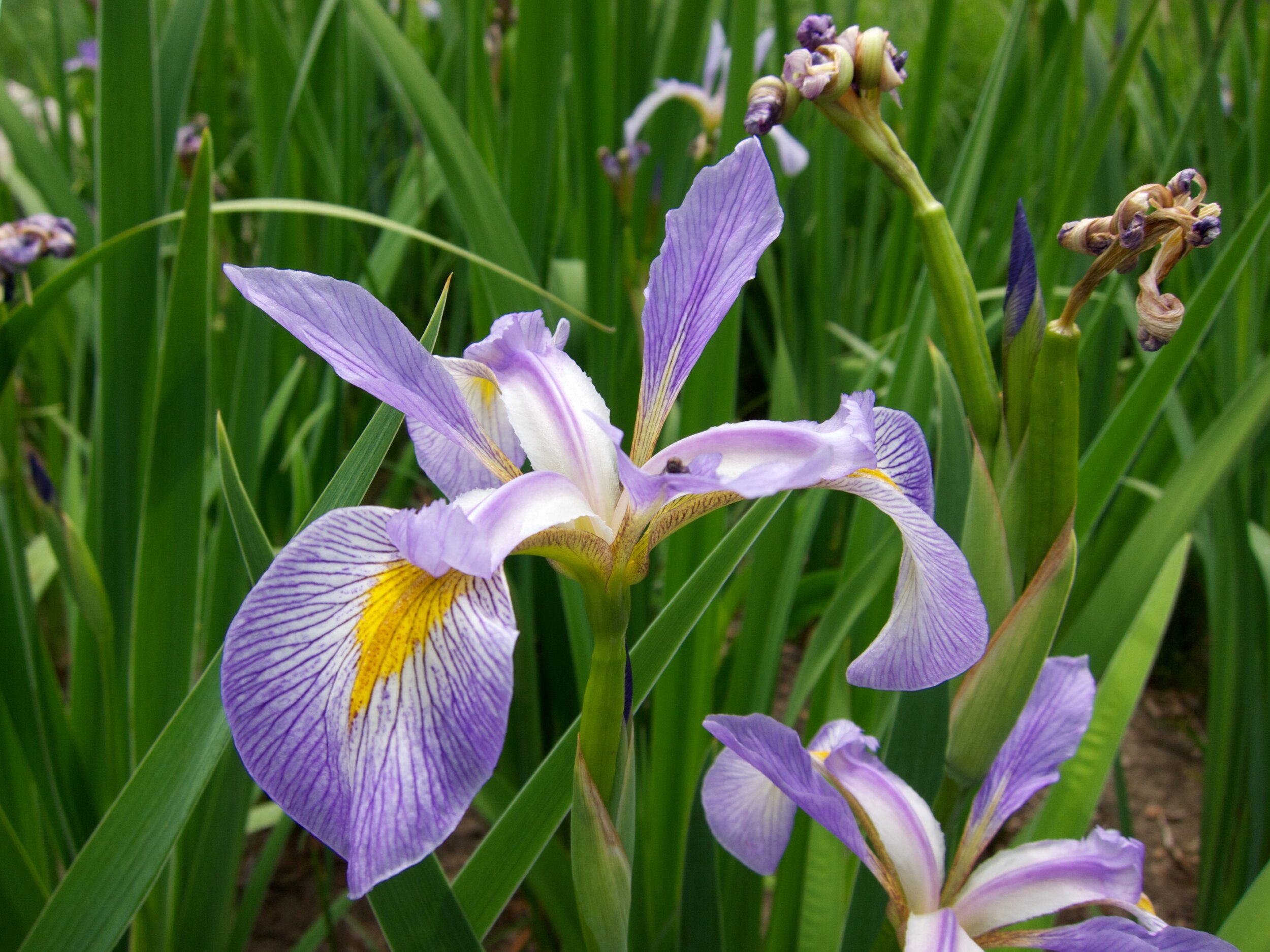
Fossils Shine Light On the History of Gall-Making Wasps

M J Richardson / Common spangle galls / CC BY-SA 2.0

M J Richardson / Common spangle galls / CC BY-SA 2.0

We can learn a lot about life on Earth from the fossil record. I am always amazed by the degree of scrutiny involved in collecting data from these preserved remains. Take, for instance, the case of gall-making wasp fossils found in western North America. A small collection of fossilized oak leaves is giving researchers insights into the evolutionary history of oaks and the gall-making wasps they host.

Oaks interact with a bewildering array of insects. Many of these are gall-making wasps in the family Cynipidae. Dozens of different wasp species can be found on a single oak tree. Female wasps lay their eggs inside developing oak tissues and the larvae release hormones and other chemicals that cause galls to form. Galls are essentially edible nursery chambers. Other than their bizarre shapes and colors, the compounds released by the wasp larvae reduce the chemical defenses of the oak and increase the relative nutrition of the tissues themselves. Often, these relationships are precise, with specific wasp species preferring specific oak species. But when did these relationships arise? Why are oaks so popular? What can fossil evidence tell us about this incredible relationship?

Photo by Beentree licensed under CC BY-SA 4.0

Photo by Beentree licensed under CC BY-SA 4.0

Though scant, the little fossil evidence of oak galls can tell us a lot. For starters, we know that gall-making wasps whose larvae produce structures similar to that of the Cynipids have been around since at least the late Cretaceous, some 100 million years ago. However, it is hard to say for sure exactly who made these galls and exactly what taxonomic affinity the host plant belongs to. More conclusive Cynipid gall fossils appear again in the Eocene and continue to pop up in the fossil record throughout the Oligocene and well into the Miocene (33 - 23 million years ago).

Miocene aged fossils are where things get a little bit more conclusive. Fossil beds located in the western United States have turned up fossilized oak leaves complete with Cynipid galls. The similarity of these galls to those of some present day species is incredible. It demonstrates that these relationships arose early on and have continued to diversify ever since. What's more, thanks to the degree of preservation in these fossil beds, researchers are able to make some greater conclusions about why gall-making wasps and oaks seem to be so intertwined.

Holotype of Antronoides cyanomontanus galls on fossilized leaves of Quercus simulata. 1) Impression of the abaxial surface of the leaf, showing the galls extending into the matrix. 2) Galls showing close association with secondary veins. 3) Gall sho…

Holotype of Antronoides cyanomontanus galls on fossilized leaves of Quercus simulata. 1) Impression of the abaxial surface of the leaf, showing the galls extending into the matrix. 2) Galls showing close association with secondary veins. 3) Gall showing the impression of rim-like base partially straddling the secondary vein. 4) Close-up of gall attached at margin extending down into the matrix. 5) Gall uncovered revealing spindle-shaped morphology.

1) Xanthoteras clavuloides galls on fossilized Quercus lobata, showing gall attached to secondary vein. Specimen in California Academy of Sciences Entomology collection, San Francisco. 2) Two galls of attached to a secondary vein showing overlap of …

1) Xanthoteras clavuloides galls on fossilized Quercus lobata, showing gall attached to secondary vein. Specimen in California Academy of Sciences Entomology collection, San Francisco. 2) Two galls of attached to a secondary vein showing overlap of their bases. Specimen in California Academy of Sciences Entomology Collection, San Francisco. 3) Three galls collected from leaf of California Quercus lobata showing clavate shape and expanded, ring-like base. 4) Gall showing the annulate or ribbed aspect of the base, which is similar to bases of Antronoides cyanomontanus and A. polygonalis. 5) Galls showing clavate shape, pilose and nonpilose surfaces, and bases.

Gall-making wasps seem to diversify at a much faster rate in xeric climates. The fossil records during this time show that mesic tree speciess were gradually being replaced by more xeric species like oaks. Wasps seem to prefer these drier environments and the thought is that such preferences have to do with disease and parasite loads.

Again, galls a large collections of nutrient-rich tissues that are low in defense compounds. Coupled with the juicy grub at their center, it stands to reason that galls make excellent sites of infection for fungi and other parasites. By living in drier habitats, it is believed that gall-making wasps are able to escape these environmental pressures that would otherwise plague them in wetter habitats. The fossil evidence appears to support this hypothesis and today we see similar patterns. White oaks are especially drought tolerant and its this group of oaks that host the highest diversity of gall-making wasps.

Photo Credits: [1] [2] [3] [4]

Further Reading: [1] [2] [3]

Meet the Blazing Stars

Liatris_aspera3.jpg

Midsummer in North America is, among other things, Liatris season. These gorgeous plants are often referred to as blazing stars or gayfeathers, which hints at the impact their flowers have on our psyche. Whether in the garden or in the wild, Liatris are a group of plants worth getting to know a bit better.

Liatris is by and large a North American genus with only a single species occurring in the Bahamas. Though we often think of Liatris as prairie plants, the center of diversity for this group is in the southeastern United States. Taxonomically speaking, Liatris are a bit of a conundrum. Something like 40 different species have been described and, where ranges overlap, many putative hybrids have been named.

Rocky Mountain blazing star (Liatris ligulistylis). Photo by Dan Mullen licensed under CC BY-NC-ND 2.0

Rocky Mountain blazing star (Liatris ligulistylis). Photo by Dan Mullen licensed under CC BY-NC-ND 2.0

Authorities on this group cite ample confusion when it comes to drawing lines between species. Much of this confusion comes from the fact that numerous variants and intergradations exist between the various species. As mentioned, hybridization is not uncommon in this genus, which complicates matters quite a bit.

Prairie blazing star (Liatris pycnostachya). Photo by Kcauley licensed under CC BY-SA 4.0

Prairie blazing star (Liatris pycnostachya). Photo by Kcauley licensed under CC BY-SA 4.0

Liatris as a whole appears to have undergone quite an adaptive radiation in North America, with species adapting to specific soils and habitat types. Take, for instance, the case of cylindrical blazing star (L. cylindracea), marsh blazing star (L. spicata), and rough blazing star (L. aspera). The ranges of these species overlap to quite a degree, however, each prefers to grow in soils of specific texture and moisture. Marsh blazing star, as you may have guessed, prefers wetter soils whereas rough blazing star enjoys drier habitats. Cylindrical blazing star seems to enjoy intermediate soil conditions, especially where soil pH is a bit higher. As such, these three species often occur in completely different habitats. However, in places like the southern shores of Lake Michigan, they find themselves growing in close quarters and as a result, a fair amount of hybridization has occurred.

Another example of confusion comes from a species commonly known as the savanna blazing star (Liatris scariosa nieuwlandii). Many different ecotypes of this plant exist and some experts don't quite know how to deal with them all. Sometimes savanna blazing star is treated as a variant of another species called the northern blazing star (Liatris scariosa var. nieuwlandii) and sometimes it is treated as its own distinct species (Liatris nieuwlandii). Until proper genetic work can be done, it is impossible to say which, if any, are correct. 

Glandular blazing star (Liatris glandulosa). Photo by Billy Bob Bain licensed under CC BY 2.0

Glandular blazing star (Liatris glandulosa). Photo by Billy Bob Bain licensed under CC BY 2.0

Taxonomic confusion aside, the various Liatris species and variants are important components of the ecology wherever they occur. Numerous insects feed upon and raise their young on the foliage and few could argue against their flowers as pollinator magnets. All Liatris produce pink to purple flowers in splendid Asteraceae fashion. Every once in a while, an aberrant form is produced that sports white flowers. Though horticulturists have capitalized on this for the garden, at least one authority claims that these white forms are much weaker than their pink flowering parents. At least one species, the pinkscale blazing star (L. elegans), produces large, filamentous white bracts that very much resemble flowers.

Check out the bracts on the pinkscale blazing star (L. elegans)!

Check out the bracts on the pinkscale blazing star (L. elegans)!

Liatris are just as interesting below as they are above. The roots, foliage, and flowers all emerge from a swollen underground stem called a corm. The formation of these corms is one reason why some Liatris species have become so popular in our gardens. It makes them extremely hardy during the dormant season. In the spring, the corm starts forming roots. At the same time, tiny preformed buds at the top of the corm begin to grow this years crop of leaves and flowers. By the end of the growing season, the corm has reached its maximum size for that year and the plant draws down the rest of its reserves to wait out the winter.

Cylindrical blazing star (Liatris cylindracea). Photo by Joshua Mayer licensed under CC BY-SA 2.0

Cylindrical blazing star (Liatris cylindracea). Photo by Joshua Mayer licensed under CC BY-SA 2.0

During this time, some species form a layer of tissue along the edge of the corm that is much darker in coloration than what was laid down earlier in the season. This has led some to suggest that aging individual Liatris is possible. Experts believe that specimens can readily reach 30 to 40 years of age or more, however, the degree to which these dark bands indicate annual growth is up for a lot of debate. Others have found no correlation with plant age. Regardless, it is safe to say that many Liatris species can live for decades if left undisturbed.

Scrub blazing star (Liatris ohlingerae). Photo by FWC Fish and Wildlife Research Institute licensed under CC BY-NC-ND 2.0

Scrub blazing star (Liatris ohlingerae). Photo by FWC Fish and Wildlife Research Institute licensed under CC BY-NC-ND 2.0

All in all, Liatris is a very special, albeit slightly confusing, group of plants. It offers a little something for everyone. What's more, their beauty is only part of the story. These are ecologically important plants that support many great insect species. As summer wears on, make sure to get out there and enjoy the Liatris in your neck of the woods. You will be happy you did!

Photo Credits: [1] [2] [3] [4] [5] [6] [7] [8]

Further Reading: [1] [2] [3]


Toxic Nectar

35523565_2235120153181504_7723861579292213248_o.jpg

I was introduced to the concept of toxic nectar thanks to a species of shrub quite familiar to anyone who has spent time in the Appalachian Mountains. Locals will tell you to never place honeybee hives near a patch of rosebay (Rhododendron maximum) for fear of so-called "mad honey." Needless to say, the concept intrigued me.

A quick internet search revealed that this is not a new phenomenon either. Humans have known about toxic nectar for thousands of years. In fact, honey made from feeding bees on species like Rhododendron luteum and R. ponticum has been used more than once during times of war. Hives containing toxic honey would be placed along known routs of Roman soldiers and, after consuming the seemingly innocuous treat, the soldiers would collapse into a stupor only to be slaughtered by armies lying in wait.

Rhododendron luteum. Photo by Chrumps licensed under CC BY 3.0

Rhododendron luteum. Photo by Chrumps licensed under CC BY 3.0

The presence of toxic nectar seems quite confusing. The primary function of nectar is to serve as a reward for pollinators after all. Why on Earth would a plant pump potentially harmful substances into its flowers?

It is worth mentioning at this point that the Rhododendrons aren't alone. A multitude of plant species produce toxic nectar. The chemicals that make them toxic, though poorly understood, vary almost as much as the plants that make them. Although there have been repeated investigations into this phenomenon, the exact reason(s) remain elusive to this day. Still, research has drummed up some interesting data and many great hypotheses aimed at explaining the patterns.

Catalpa nectar has been shown to deter some ants and butterflies but not large bees. Photo by Le.Loup.Gris licensed under CC BY-SA 3.0

Catalpa nectar has been shown to deter some ants and butterflies but not large bees. Photo by Le.Loup.Gris licensed under CC BY-SA 3.0

The earliest investigations into toxic nectar gave birth to the pollinator fidelity hypothesis. Researchers realized that meany bees appear to be less sensitive to alkaloids in nectar than are some Lepidopterans. This led to speculation that perhaps some plants pump toxic compounds into their nectar to deter inefficient pollinators, leading to more specialization among pollinating insects that can handle the toxins.

Another hypothesis is the nectar robber hypothesis. This hypothesis is quite similar to the pollinator fidelity hypothesis except that it extends to all organisms that could potentially rob nectar from a flower without providing any pollination services. As such, it is a matter of plant defense.

The nectar of Cyrilla racemiflora is thought to be toxic to some bees. Photo by Koala:Bear licensed under CC BY-SA 2.0

The nectar of Cyrilla racemiflora is thought to be toxic to some bees. Photo by Koala:Bear licensed under CC BY-SA 2.0

Others feel that toxic nectar may be less about pollinators or nectar robbers and more about microbial activity. Sugary nectar can be a breeding ground for microbes and it is possible that plants pump toxic compounds into their nectar to keep it "fresh." If this is the case, the antimicrobial benefits could outweigh the cost to pollinators that may be harmed or even deterred by the toxic compounds.

Finally, it could be that toxic nectar may have no benefit to the plant whatsoever. Perhaps toxic nectar is simply the result of selection for defense compounds elsewhere in the plant and therefore is expressed in the nectar as a result of pleiotropy. If this is the case then toxic nectar might not be under as strong selection pressures as is overall defense against herbivores. If so, the plants may not be able to control which compounds eventually end up in their nectar. Provided defense against herbivores outweighs any costs imposed by toxic nectar then plants may not have the ability to evolve away from such traits.

Where Spathodea campanulata is invasive, its nectar causes increased mortality in native bee hives. Photo by mauro halpern licensed under CC BY 2.0

Where Spathodea campanulata is invasive, its nectar causes increased mortality in native bee hives. Photo by mauro halpern licensed under CC BY 2.0

So, where does the science land us with these hypotheses? Do the data support any of these theories? This is where things get cloudy. Despite plenty of interest, evidence in support of the various hypotheses is scant. Some experiments have shown that indeed, when given a choice, some bees prefer non-toxic to toxic nectar. Also, toxic nectar appears to dissuade some ants from visiting flowers, however, just as many experiments have demonstrated no discernible effect on bees or ants. What's more, at least one investigation found that the amount of toxic compounds within the nectar of certain species varies significantly from population to population. What this means for pollination is anyone's' guess.

It is worth noting that most of the pollination-related hypotheses about toxic nectar have been tested using honeybees. Because they are generalist pollinators, there could be something to be said about toxic nectar deterring generalist pollinators in favor of specialist pollinators. Still, these experiments have largely been done in regions where honeybees are not native and therefore do not represent natural conditions.

Simply put, it is still too early to say whether toxic nectar is adaptive or not. It could very well be that it does not impose enough of a negative effect on plant fitness to evolve away from. More work is certainly needed. So, if you are someone looking for an excellent thesis project, here is a great opportunity. In the mean time, do yourself a favor and don't eat any mad honey.

Photo Credits: [1] [2] [3] [4]

Further Reading: [1] [2] [3] [4] [5] [6]